The medulla oblongata, commonly known as the medulla, stands as a critical component of the brainstem, connecting the spinal cord with higher brain structures. As the most inferior region of the brainstem, the medulla plays a vital role in regulating essential autonomic functions necessary for our survival, including breathing, heartbeat, and digestion. This article delves into the intricacies of the medulla, exploring its external and internal anatomy, along with its crucial blood supply. Understanding the medulla’s anatomy and functions is key to appreciating how it orchestrates an array of automatic and voluntary actions that sustain human life and well-being.
External Anatomy of the Medulla oblongata
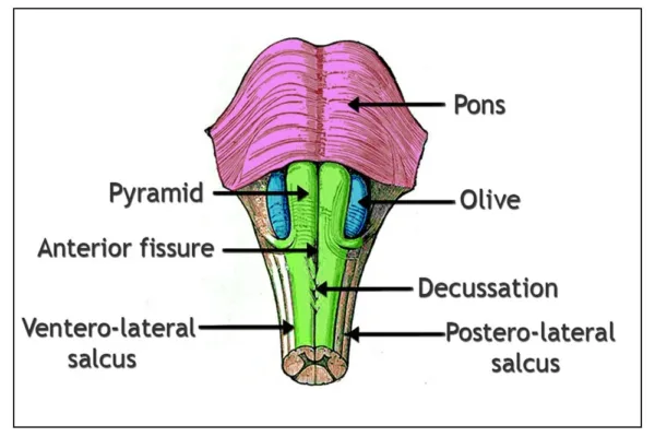
- The medulla is conical in shape, tapering down as it extends inferiorly.
- It is approximately 3 cm long and 2 cm wide at its widest point.
- The superior margin of the medulla is located at the junction with the pons, and the inferior margin is marked by the origin of the first pair of cervical spinal nerves.
- On the anterior surface, three fissures are visible: the anterior median fissure, ventrolateral sulcus, and posterolateral sulcus.
- Structures on the anterior surface include the pyramids (corticospinal tracts responsible for motor control), the olives (containing nerve fibers connecting the cerebellum and pons with the medulla), and five cranial nerves (CN VI, CN XII, CN IX, CN X, and CN XI).
- The posterior surface is relatively obscured by the cerebellum, but the posterior median sulcus and the dorsal column-medial lemniscus pathway (fasciculus gracilis and fasciculus cuneatus) can be observed.
Internal Anatomy of the Medulla oblongata
- The internal anatomy can be observed in cross sections at three levels: the decussation of the pyramids, the decussation of the medial lemnisci, and the level of the olives.
- At the level of the decussation of the pyramids, the lateral corticospinal tracts are formed by 75% of motor fibers crossing diagonally and posteriorly.
- The posterior white matter contains the fasciculus gracilis and fasciculus cuneatus, carrying sensory information from the spinal cord. The trigeminal nucleus and tracts are also visible.
- At the level of the decussation of the medial lemnisci, sensory decussation occurs, and the internal arcuate fibers form the medial lemniscus. The trigeminal nucleus and spinal tract, tectospinal tract, and vestibular nuclei can be observed.
- At the level of the olives, the central canal expands into the fourth ventricle, making this region the “open part” of the medulla. Cranial nerve nuclei, such as the hypoglossal nucleus and nucleus ambiguous, are visible, as well as the lateral spinothalamic tract and nucleus of the tractus solitarius.
Vasculature:
- Medulla receives blood supply from multiple arteries, ensuring its proper functioning and vital role in regulating essential bodily functions.
- The anterior spinal artery is one of the key suppliers, providing blood to the ventral portion of the medulla, including the pyramids responsible for motor control.
- Another significant artery is the posterior spinal artery, which supplies blood to the posterior half of the medulla, supporting sensory functions and information processing.
- The posterior inferior cerebellar artery is also involved in the blood supply to the medulla, contributing to various regions and maintaining its overall health.
- Equally important is the anterior inferior cerebellar artery, ensuring blood flow to specific areas of the medulla, enabling its diverse functions.
The Role of Vertebral Arteries:
Vertebral arteries play a crucial role in delivering blood to the medulla, sustaining its connection with the spinal cord and higher brain structures. The blood supply to the medulla varies depending on the level being viewed, with each artery catering to specific regions and nuclei for optimized performance. Adequate blood circulation in the medulla is essential for maintaining autonomic functions such as breathing, heartbeat, and digestion, making these arteries vital for overall well-being. The complex network of arteries ensures a constant and sufficient supply of oxygen and nutrients, supporting the medulla’s role in regulating critical bodily processes. A healthy blood supply to the medulla is essential for coordinating the integration of sensory and motor information, contributing to proper homeostasis and overall brain health.
Conclusion:
In summary, the medulla oblongata is a crucial part of the brainstem that regulates essential autonomic functions. Its external and internal anatomy reveals distinct structures and nuclei responsible for motor and sensory functions. Proper blood supply is vital for its normal functioning and maintenance of overall brain health. Understanding the anatomy of the medulla is essential for comprehending its role in maintaining various bodily functions.
Functions of Medulla Oblongata:
The medulla oblongata is a critical structure within the brainstem that plays a central role in controlling numerous bodily functions. It houses the nuclei of the lower four cranial nerves, which are crucial for various sensory and motor functions in the head and neck region.
Glossopharyngeal Nerve (CN IX):
The medulla controls functions via the glossopharyngeal nerve, including:
- Coordination of Swallowing: Safely transports food and liquids from mouth to esophagus.
- Salivation Regulation: Aids digestion and maintains oral health by controlling saliva production.
- Visceral Sensation Conveyance: Transmits sensory information, including taste perception, from oral cavity and throat.
Vagus Nerve (CN X):
The medulla regulates various bodily functions through the vagus nerve, such as:
• Parasympathetic Supply: Controlling the parasympathetic division of the autonomic nervous system, which is responsible for the “rest and digest” response. This includes regulating heart rate, respiratory rate, and digestive processes.
• Gland Secretion Control: Influencing secretions from various glands, including those in the head, thorax, and abdomen.
• Peristalsis: Coordinating the rhythmic contractions of smooth muscles in the gastrointestinal tract to facilitate food movement.
• Phonation: Controlling the muscles responsible for speech production.
• Taste Sensation: Relaying taste information from the back of the throat and the digestive tract.
Accessory Nerve (CN XI):
The medulla plays a role in controlling movements of the head and shoulders via the accessory nerve, particularly during phonation and head movements.
Hypoglossal Nerve (CN XII):
The medulla regulates movements of the tongue, speech articulation, and swallowing through the hypoglossal nerve.
Respiratory Center:
The medulla oblongata contains a complex group of nuclei that form the respiratory center. This center is responsible for regulating breathing, ensuring that oxygen is supplied to the body and carbon dioxide is eliminated.
It consists of three parts: the dorsal respiratory group, ventral respiratory group, and pneumotaxic center.
• Dorsal Respiratory Group: Responsible for initiating inhalation (inspiration) by stimulating the contraction of the diaphragm and intercostal muscles. It receives information from peripheral chemoreceptors regarding blood oxygen levels.
• Ventral Respiratory Group: Activated during increased pulmonary ventilation, it stimulates accessory respiratory muscles to increase respiratory effort.
• Pneumotaxic Center: Located in the pons, this center regulates the respiratory cycle and the duration of inspiration, helping control breathing patterns.
Vasomotor Center:
The medulla’s vasomotor center plays a vital role in regulating arterial blood pressure. It receives signals from baroreceptors in the aortic body, which provide information about blood pressure. Based on this information, the vasomotor center initiates autonomic responses to influence heart rate and the diameter of blood vessels throughout the body.
Integration of Cardiovascular and Respiratory Functions: Research suggests that the medulla oblongata integrates cardiovascular and respiratory functions within specific regions. Rostral ventral lateral medulla (RVLM) contains excitatory neurons that provide information to pre-sympathetic neurons in the spinal cord, maintaining baseline arterial pressure. Ventral respiratory column, also within the RVLM, controls respiratory rhythm and pattern, influencing the oscillating respiratory pattern for optimal oxygen perfusion in tissues. Medulla’s various nuclei and pathways also coordinate with the cardiovascular system’s regulation to maintain homeostasis.
Other Medullary Nuclei and Functions:
Apart from the cranial nerve nuclei and respiratory and vasomotor centers, the medulla oblongata houses other important nuclei with specific functions:
• Nucleus of the Solitary Tract (NTS): This nucleus processes and coordinates visceral afferent information related to cardiorespiratory functions, including signals from peripheral chemoreceptors, pulmonary stretch receptors, and baroreceptors.
• Area Postrema: Located on the dorsal surface of the medulla, it serves as the vomiting center, detecting emetic toxins in the blood and cerebrospinal fluid.
• Spinal Trigeminal Nucleus: Involved in processing sensory information from the face, including pain, temperature, and touch sensation.
• Inferior Olivary Nuclei: Important for transmitting proprioception, muscle tension, and motor intention information to the cerebellum.
• Reticular Formation: A complex network found throughout the brainstem, involved in autonomic regulation of respiration, heart rate, and blood pressure.
Conclusion:
In summary, the medulla oblongata is a vital brainstem region that controls a wide range of essential bodily functions. Its integration of cranial nerves, respiratory and cardiovascular centers, and other nuclei allows for the coordination of various processes critical to sustaining life. Without the medulla, many automatic and voluntary actions we take for granted would be compromised, highlighting the crucial role this region plays in maintaining human homeostasis and well-being. Understanding the anatomy and functions of the medulla sheds light on its significance in our overall health and underscores the importance of protecting and nurturing this essential brain structure.
Learn more
References
- Bae, Y. J., Petrus, D. S., & Gatto, G. J. (2019). Anatomy, Brainstem. In StatPearls [Internet]. StatPearls Publishing.
- Benarroch, E. E. (2015). Brainstem: anatomy and function. Handbook of Clinical Neurology, 128, 1-13.
- Carpenter, M. B., & Sutin, J. (1983). Human neuroanatomy (8th ed.). Williams & Wilkins.
- Kiernan, J. A., & Barr, M. L. (2013). Barr’s the human nervous system: An anatomical viewpoint (10th ed.). Lippincott Williams & Wilkins.
- Koeppen, A. H., & Stanton, B. A. (2017). Berne and Levy Physiology (7th ed.). Elsevier.
- Martin, J. H. (2019). Neuroanatomy: Text and Atlas (5th ed.). McGraw-Hill Education.
- Mendoza, J. E., & Foundas, A. L. (2018). Clinical neuroanatomy: A neurobehavioral approach (3rd ed.). Springer.
- Nieuwenhuys, R., Voogd, J., & van Huijzen, C. (2008). The human central nervous system (4th ed.). Springer.
- Reis, D. J., & Doba, N. (1983). Brainstem cardiovascular centers. In Handbook of Physiology. The Cardiovascular System. American Physiological Society.
- Standring, S. (Ed.). (2016). Gray’s Anatomy: The Anatomical Basis of Clinical Practice (41st ed.). Elsevier.