Define cell junction
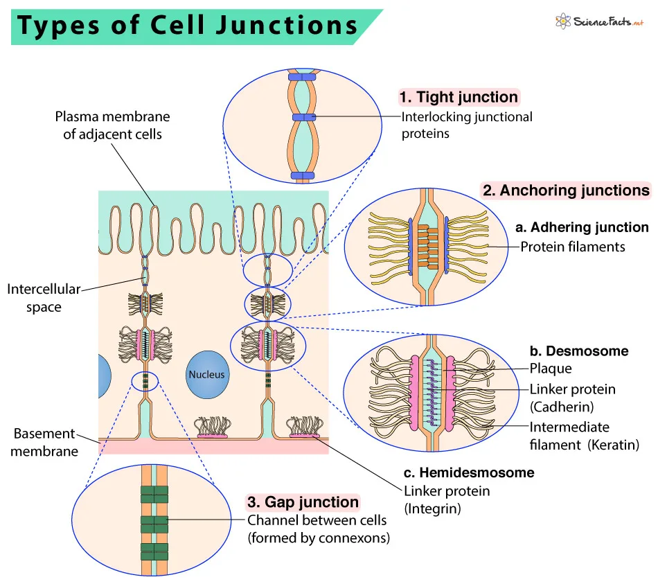
Cell junctions are specialized structures that link cells to one another and to the extracellular matrix (ECM) that surrounds them. They are essential for tissue integrity and function because they promote cell communication and coordination. Tight junctions, adherens junctions, desmosomes, and gap junctions are all different types of cell junctions, each having their own structural and functional qualities.
Tight junctions
- Tight junctions, which create a continuous seal between neighboring cells, are the most apical (closest to the surface) of the cell junctions present in epithelial and endothelial tissues.
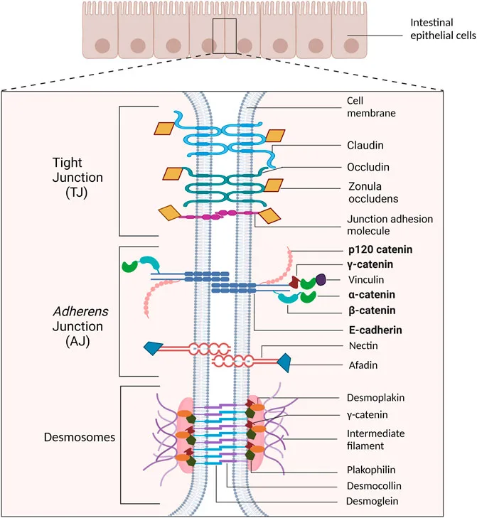
- Tight junctions are composed of transmembrane proteins known as claudins, occludins, and junctional adhesion molecules (JAMs), which are attached to the cytoskeleton through a scaffold protein complex.
- Tight junctions act as gatekeepers, controlling the movement of chemicals and ions via the paracellular space (the area between neighboring cells).
- The expression and distribution of tight junction proteins, as well as the activity of numerous signaling pathways, influence the seal’s tightness.
- Tight junctions are critical for tissue polarity and inhibiting lateral membrane protein diffusion between distinct regions of the plasma membrane.
- Tight junction disruption can result in increased paracellular permeability, which can contribute to a variety of disorders such as inflammatory bowel disease, celiac disease, and cancer.
- Tight junctions also play a role in the formation and maintenance of the blood-brain barrier, which controls the transit of chemicals and ions between the circulation and the brain parenchyma.

Types of tight junction
Tight junctions are cell-cell junctions that play an important function in controlling epithelial and endothelial paracellular permeability. Tight junctions are classified according to their composition and function. Here are a few examples of tight junctions:
- Occludin-based tight junctions: These tight junctions are made up of the transmembrane protein occludin and related proteins like as ZO-1, ZO-2, and ZO-3. They are typically present in epithelial and endothelial cells.
- Claudin-based tight junctions: These tight junctions are made up of the transmembrane protein claudin and related proteins like as ZO-1, ZO-2, and ZO-3. They are the most common form of tight junction and are seen in a variety of tissues.
- Junctional adhesion molecule (JAM)-based tight junctions: These tight junctions are made up of JAMs and related proteins including ZO-1, ZO-2, and ZO-3. They are mostly present in endothelial cells and leukocytes.
- Tricellulin-based tight junctions: These tight junctions are located at tricellular contacts, where three cells come together. They are made up of the transmembrane protein tricellulin and related proteins such as MAGI-1 and MPP5.
- ESAM-based tight junctions: These tight junctions are made up of endothelial cell-selective adhesion molecule (ESAM) and related proteins including ZO-1, ZO-2, and ZO-3. They are mostly present in endothelial cells.
- Clotting factor XIIIa (FXIIIa)-dependent tight junctions: These tight junctions are generated by the crosslinking of occludin and claudin by FXIIIa, a transglutaminase enzyme. They are present in epithelial cells.
In all, tight junction composition varies based on tissue type and physiological environment. Tight junctions are essential for tissue integrity and barrier function, and abnormalities in their structure or function can lead to illnesses including inflammatory bowel disease and cancer.
Adherens junction
Adherens junctions are cell-cell junctions which are essential for tissue integrity, cell-cell adhesion, and signal transmission. Here are some items to consider to better comprehend adherens junctions:
- Cadherin, a transmembrane glycoprotein, forms adherens junctions by interacting with other cadherin molecules on neighboring cells via homophilic binding.
- Cadherin is attached to the actin cytoskeleton using a complex of proteins, which includes beta-catenin along with alpha-catenin, which are also essential to junction stability.
- Adherens junctions are dynamic structures that may build and deconstruct quickly when subjected to different signaling routes and physical pressures.
- Adherens junctions may generate linear belts surrounding the apical tips of epithelial cells, that are necessary for tissue polarization and barrier functionality.
- Adherens junctions can additionally form punctate patches at mesenchymal cell-cell interactions, in which they govern cell adhesion and migration.
- Adherens junctions are controlled by a variety of signaling pathways, such as the Wnt/beta-catenin pathway, which regulates junction integrity and renewal.
- Adherens junctions are vital for tissue growth and homeostasis, and abnormalities in their functioning have been linked to a variety of illnesses such as cancers and developmental problems.
- Adherens junctions are also important in mechanotransduction, as they convey mechanical forces between cells and control stiffness of tissue and tensions.
- Adherens junctions differ from other types of cell-cell junctions, such as tight junctions and gap junctions, in their structure and function.

Types of adherens junction
Adherens junctions are a form of cell junction that links neighboring cells. Adhesive junctions are classified into two types:
- Cadherin-based Adherens Junctions: Cadherin-based adhesion junctions are generated largely by the transmembrane protein cadherin, that links to cadherins on nearby cells, connecting them. These junctions may be observed in a variety of tissue & are vital for tissue integrity and cell adhesion.
- Desmosome-based Adherens Junctions: Desmosomes, complex structures of proteins that tether intermediate filaments to the cell membrane, build these junctions. Desmosomes are most prevalent in tissues subjected to mechanical stress, such as the skin and heart, and they serve to avoid tearing and to preserve tissue strength.
Gap junction
Gap junctions are intercellular channels that enable direct communication and the exchange of ions, tiny molecules, and electrical impulses between neighboring cells. Here are some crucial aspects to consider when learning about gap junctions:
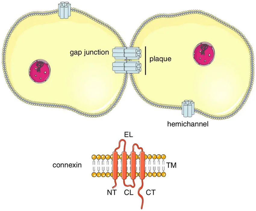
- Gap junctions are made by a set of proteins named connexins that are organized in a hexagonal configuration that creates a pore-like structure called a connexon.
- Each connexon is made up of six connexin subunits, and two connexons from nearby cells join together to form a gap junction channel.
- Gap junctions exist in a variety of tissues, including the brain, heart, liver, and epithelial tissues.
- Gap junctions facilitate the exchange of tiny molecules (1 kDa) among cells, which includes as ions, carbohydrates, and amino acids.
- Gap junctions also facilitate the interchange of electrical impulses, such as action potentials, between cells in excitable organs such as the heart and brain.
- Gap junctions are important for coordinating the activity of groups of cells, such as cardiac muscle cells or neurons.
- The opening and shutting of gap junctions can be modulated by a variety of variables like as pH, calcium levels, and phosphorylation.
- Mutations or alterations in connexin expression can potentially impact gap junctions, resulting in illnesses such as deafness, cataracts, and many neurological problems.
- Gap junctions vary from other types of cell-cell junctions, such as adherens junctions and tight junctions, in their structure and function.
- Electron microscopy, dye transfer experiments, and patch-clamp recordings may all be used to view and study gap junctions.
Types of Gap junction
Gap junctions are specialized intercellular channels that allow ions, tiny molecules, and electrical impulses to be exchanged directly between nearby cells. Gap junctions are classified into various categories, including:
- Connexons: These are the basic building blocks of gap junctions and are made up of six connexin proteins that join together to create a hexameric structure. There are about 20 distinct kinds of connexins, each one having its own set of characteristics and locations across various tissues.
- Hemi-channels: These are half of a connexon that can operate autonomously of the entire hexameric arrangement. Hemi-channels are present on the surface of individual cells and can interact with external substances.
- Electrical synapses: These are specially designed gap junctions that allow for the direct transmission of electrical impulses between neurons. Electrical synapses are critical in controlling the timing of neuronal activity and the development of synchronous firing patterns.
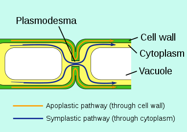
- Plasmodesmata: They are distinctive gap junctions present in plant cells that allow chemicals and tiny particles to be exchanged directly between neighboring cells. Plasmodesmata serve as vital for transporting nutrients and signaling chemicals between plant tissues.
Desmosomes

- Desmosomes are specialized cell-cell junctions which provide mechanical strength to mechanically stressed tissues like skin and heart muscle.
- Desmosomes are composed of transmembrane proteins that are known as desmogleins and desmocollins, which interact in a calcium-dependent way to produce desmosomal cadherin complex.
- Cadherin complexes on the desmosomal half of the plasma membrane link to intermediate filaments, that are attached to the cytoplasmic plaque and the cytosolic side of the plasma membrane.
- Intermediate filaments, which are made up of keratin in epithelial cells and desmin in muscle cells, offer the tensile strength required for tissues to tolerate physical strain.
- The desmosomal plaque contains numerous additional proteins, such as as plakoglobin, plakophilin, and desmoplakin, which anchor intermediate filaments to the cytoplasmic side of the plasma membrane.
- Desmosomes are dynamic structures that can rearrange in response to mechanical stress or signaling pathways.
- Desmosomal protein mutations can cause a number of illnesses, including skin blistering disorders, arrhythmogenic cardiomyopathy, and autoimmune diseases such as pemphigus vulgaris.
Plasmodesmata
Plasmodesmata are small channels generated by cell walls that link neighboring plant cells. Here are some crucial points concerning the anatomy and functioning of plasmodesmata:
Structure of Plasmodesmata
1. Plasmodesmata are cytoplasmic channels enclosed by a cell wall and bordered with plasma membrane.
2. They are typically seen at the cell wall corners where three or more cells meet.
3. Plasmodesta vary in size and structure, although they are generally 50-60 nm in diameter.
4. The channels are bordered by a lipid bilayer plasma membrane that is contiguous with the membranes of the two neighboring cells.
5. A thick cytoplasmic sleeve that is contiguous with the cytoplasm of the two neighboring cells fills the center section of the plasmodesmata.
Function of Plasmodesmata
1. Plasmodesmata allow molecules to interact between neighbouring plant cells.
2. They promote cell communication and the movement of substances like as hormones, carbohydrates, amino acids, and ions.
3. Plasmodesmata also allow organelles, such as chloroplasts, to travel between cells.
4. They are essential for plant development and growth, as well as their reactions to environmental stimuli.
5. Plasmodesmata are also involved in plant defense processes, allowing for the transmission of signals and chemicals that can stimulate immunological responses in nearby cells.
Functions of Cell junctions
Cell junctions are specialized structures that join neighboring cells and play an important role in tissue growth, homeostasis, and function. Here are some of the most important roles of cell junctions:
- Cell adhesion: Cell junctions assist to connect neighbouring cells, forming the structural underpinning for tissue and organs.
- Barrier function: Tight junctions, for example, provide a barrier that stops molecules and ions from passing between cells, aiding in tissue integrity and regulating the transport of substances among epithelial layers.
- Communication: Gap junctions enable direct interchange of chemicals, ions, and electrical impulses between cells, allowing for more coordinated responses to environmental changes and stimuli.
- Signaling: Cell junctions can function as signaling channels, conveying messages from the extracellular matrix to the cell’s interior and assisting in controlling processes of cellular activity such as development, differentiation, proliferation, and death.
- Mechanical support: Some cell junctions, such as desmosomes, bind intermediate filaments to the cell membrane, providing strong mechanical support for tissues that endure mechanical stress, such as the skin and heart.
- Development: Cell junctions play an important role in tissue development, including cell migration, tissue creation, and organogenesis.
- Disease: Cell junction dysfunction has been associated to a number of illnesses, including cancer, skin problems, and cardiovascular disease.