Centrioles are among the most sophisticated and geometrically precise subcellular structures in the animal kingdom. Traditionally viewed as the “engines” of cell division, modern research has unveiled their complex roles in sensory function, embryogenesis, and the mechanics of human fertility. This article explores the structure, function, and clinical significance of centrioles, with a specific focus on the groundbreaking discovery of centrosome remodeling in human sperm.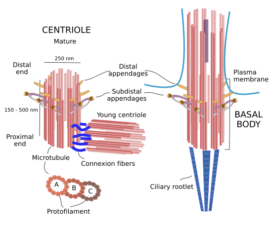
1. Structure and Dimensions: The Microtubule Barrel
The centriole is a barrel-shaped organelle located within the centrosome, the primary microtubule-organizing center (MTOC) of the cell. Physically, centrioles are constructed from specialized proteins called microtubules.
Geometry: In animal cells, a mature centriole typically consists of nine triplet microtubules arranged in a pinwheel symmetry.
Dimensions: On average, a centriole measures 200 nm in diameter and 500 nm in length.
Formation: While centrioles do not contain their own DNA, they are self-duplicating structures. They utilize “massules”—proteinaceous nucleating centers—to facilitate the assembly of new centrioles during the cell cycle.
2. Fundamental Functions in Cellular Dynamics
Centrioles are not merely structural anchors; they are dynamic regulators of cellular architecture and communication.
Cell Division (Mitosis and Meiosis)
The centrosome is the architect of the microtubular cytoskeleton. During the anaphase stage of mitosis, centrioles facilitate the formation of spindle fibers. These fibers are responsible for pulling sister chromatids apart toward opposite poles of the cell. Without functional centrioles, the accurate segregation of chromosomes would fail, leading to genomic instability.
Ciliogenesis and Sensory Function
The most evolutionarily conserved function of the centriole is the formation of cilia and flagella. In a process known as ciliogenesis, the centriole migrates to the cell surface to act as a basal body. These structures are vital for:
Cell Motility: Enabling cells to move through fluid environments.
Lung Function: Cilia in the respiratory tract sweep away mucus and debris.
Sensory Perception: Cilia are essential for vision (photoreceptors) and hearing.
Cell Differentiation
Centrioles govern asymmetrical cell division. By polarizing the cell architecture, they ensure that daughter cells receive different molecular signals, which is a fundamental requirement for the development of diverse tissue types from stem cells.
3. Centrioles in Human Reproduction: The “Sperm Aster”
A profound area of study in reproductive biology is the role of the centriole in fertilization. While the human oocyte (egg) lacks centrioles—containing only Pericentriolar Material (PCM) dissolved in the cytosol—the sperm contributes two remodeled centrioles to the zygote.
Centrosome Remodeling in Spermiogenesis
During the development of sperm (spermiogenesis), the centrosome undergoes significant remodeling. This process involves:
Protein Enrichment: The loss of certain centrosomal proteins and the enrichment of others.
Formation of the Atypical Centriole: The Distal Centriole (DC) transforms into an atypical structure, often featuring doublet microtubules instead of the standard triplets.
The Sperm Aster: Upon fertilization, the sperm centrioles recruit the maternal PCM from the egg’s cytoplasm to form the functional zygote centrosome. This structure creates the “sperm aster,” which facilitates the migration of the male and female pronuclei toward each other for fusion (karyogamy).
4. Clinical Significance and Pathology
Because centrioles are central to so many biological pathways, their dysfunction is linked to a variety of severe medical conditions:
Oncology: Defects in centriole number or structure can lead to multipolar spindles, resulting in aneuploidy and the progression of cancer.
Developmental Disorders: Mutations affecting centriole biogenesis are primary causes of microcephaly (reduced brain size) and certain forms of blindness.
Male Infertility: Insights into the atypical centriole have opened new avenues for diagnosing “unexplained” male infertility. If the sperm’s remodeled DC fails to participate in spindle pole formation or pronuclei congregation, the embryo cannot develop.
5. Conclusion: A New Diagnostic Frontier
The discovery that human sperm contains two remodeled, atypical centrioles has challenged long-standing biological dogmas. These structures are now recognized as critical signals for the differential fates of the zygote’s daughter cells and the successful start of embryonic life.
Understanding the molecular signatures of these remodeled centrioles provides innovative paths for diagnostic and therapeutic strategies in reproductive medicine. By identifying defects in sperm centrosome remodeling, clinicians can better predict and treat early embryo developmental failures.
Key Takeaways for Researchers
Structure: 9×3 microtubule arrangement; 200nm x 500nm.
Fertilization: Maternal PCM is recruited by paternal centrioles.
Spermiogenesis: Distal centrioles become “atypical” (doublets) to aid planar movement and signaling.
Pathology: Linked to ciliopathies, cancer, and infertility.
References
Avidor-Reiss, T., & Fishman, E. (2019). It takes two (centrioles) to tango. Reproduction, 157(2), R33-R51. doi: 10.1530/rep-18-0350.
Image Source Attribution: mmegias.webs.uvigo.es