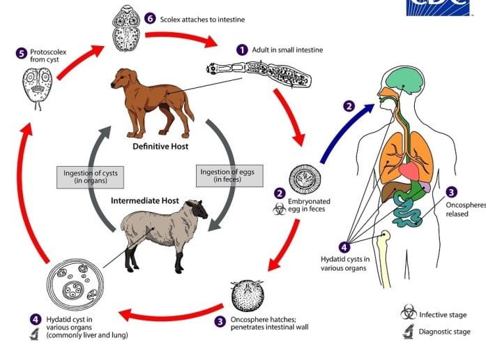
Echinococcosis is a parasitic disease caused by infection with tiny tapeworms of the genus Echinococcus. Humans are accidental hosts, contracting the disease through ingestion of the larval (egg) stage of Echinococcus granulosus. This tapeworm primarily infects dogs (the definitive hosts) and various intermediate hosts, including sheep, cattle, goats, and pigs. Dogs acquire the parasite by eating the organs of infected animals that contain hydatid cysts, which then develop into adult tapeworms in the dog. The adult tapeworms shed eggs in the feces, which contaminate the ground. Intermediate hosts ingest these eggs from contaminated environments.
Phylum: Platyhelminthes
Class: Cestoda
Genus: Echinococcus
Species: E. granulosus
Habitat and Geographical Distribution
- Adult form: Resides in the intestine of the definitive host (dog).
- Larval form: Found in the liver and lungs of humans.
Echinococcosis is distributed worldwide but is particularly prevalent in cattle-rearing countries where dogs can ingest the organs of infected animals.
Life Cycle of E. granulosus
- Adult Tapeworm: Measures 2–7 mm and lives in the small intestine of the definitive host (dog). Gravid proglottids release eggs that are immediately infectious.
- Intermediate Host: Eggs are ingested by intermediate hosts, hatch in the small intestine, and release six-hooked oncospheres. These penetrate the intestinal wall and migrate through the circulatory system to various organs, especially the liver and lungs.
- Hydatid Cyst Formation: In the liver or lungs, the oncosphere develops into a thick-walled hydatid cyst that enlarges over time. Inside the cyst, protoscolices and daughter cysts develop.
- Reinfection: The definitive host becomes infected by ingesting cyst-containing organs from an intermediate host. Protoscolices evaginate, attach to the intestinal mucosa, and mature into adult tapeworms within 32 to 80 days.
- Human Infection: Humans, as aberrant or accidental intermediate hosts, ingest eggs that release oncospheres in the intestine. Hydatid cysts develop in various organs, and if cysts rupture, protoscolices may spread to other sites, causing secondary echinococcosis.
Clinical Manifestations
- Cystic Echinococcosis: Often asymptomatic until cysts grow large enough to cause discomfort, pain, nausea, or vomiting.
- Cyst Growth: Cysts can take years to mature, with symptom appearance depending on cyst location.
- Common Locations: Liver and lungs; can also be found in the spleen, kidneys, heart, bone, and central nervous system (including brain and eyes).
- Cyst Rupture: Can lead to anaphylactic reactions or even death due to the release of cystic fluid.
Laboratory Diagnosis
- Clinical History: A cyst-like mass in a person with exposure to sheepdogs in endemic areas suggests cystic echinococcosis.
- Imaging Techniques: CT scans, ultrasonography, and MRIs are used to detect cysts.
- Serologic Tests: Once a cyst is detected, serologic tests (e.g., IHA, ELISA) confirm the diagnosis.
Treatment, Prevention, and Control
- Treatment: Surgery is the most effective method to remove cysts and can lead to a complete cure. Some inactive cysts may resolve without treatment.
- Prevention:
- Prevent dogs from eating carcasses of infected sheep.
- Control stray dog populations.
- Restrict home slaughter of sheep and other livestock.
- Avoid consuming food or water contaminated with dog fecal matter.
- Wash hands with soap and warm water after handling dogs and before eating.
- Educate children on the importance of hand hygiene to prevent infection.