The human brain is often described as the most complex object in the known universe. Weighing roughly three pounds, this jelly-like mass of fat and protein serves as the command center for the entire body. It is the seat of intelligence, the interpreter of senses, the initiator of body movement, and the controller of behavior.
Understanding the brain’s intricate anatomy is not just for neuroscientists; it is essential for anyone interested in how we perceive the world, store memories, and maintain life-sustaining rhythms. In this guide, we will break down the four primary regions of the brain: the cerebrum, the diencephalon, the cerebellum, and the brainstem, along with the protective systems that keep this vital organ safe.
1. The Cerebrum: The Seat of Higher Intelligence
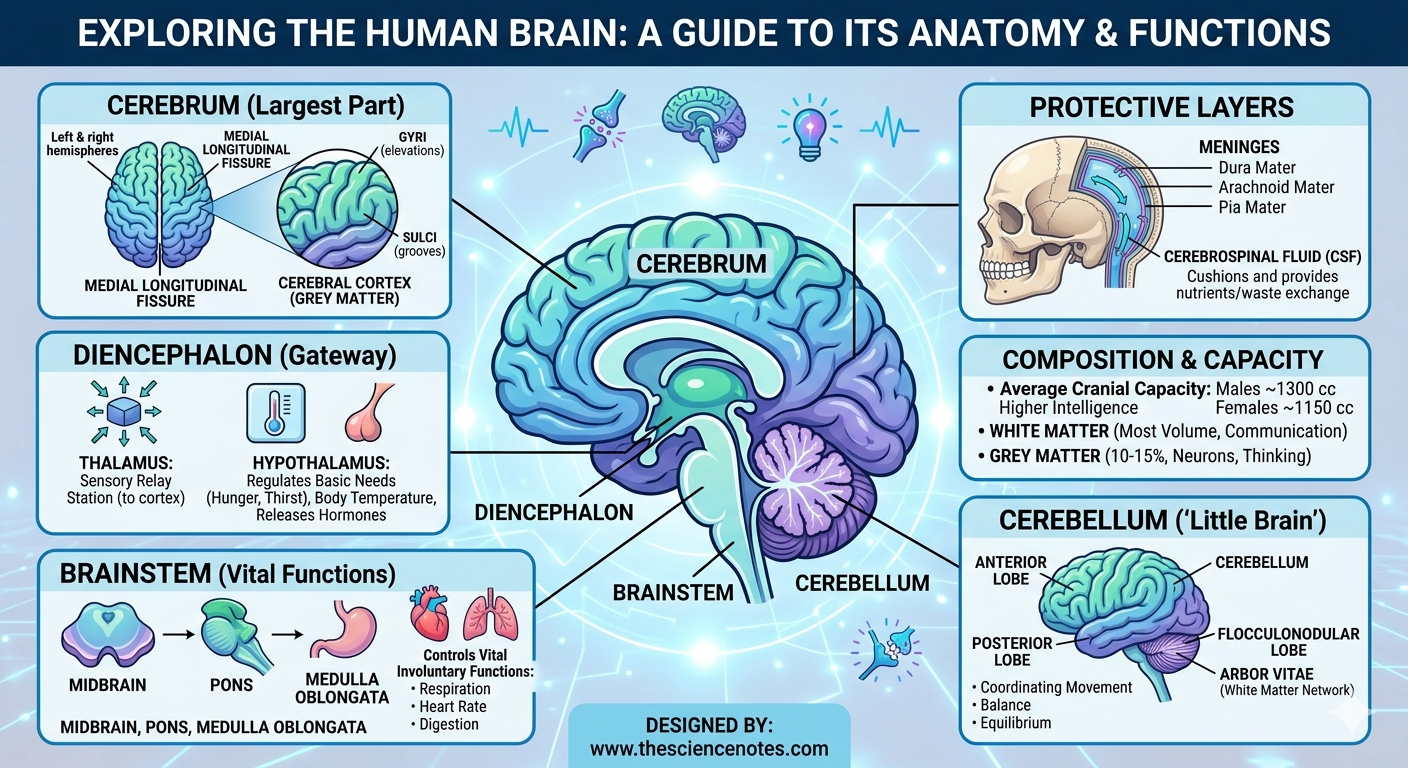
The cerebrum is the largest section of the brain, accounting for approximately 85% of its total weight. It is responsible for the functions that make us uniquely human, including conscious thought, planning, language, and sensory processing.
Hemispheres and Fissures
The cerebrum is divided into two distinct halves: the left and right hemispheres. These are separated by a deep groove known as the medial longitudinal fissure. While the two halves look like mirror images, they often specialize in different tasks—a concept known as lateralization. They communicate through a thick bundle of nerve fibers called the corpus callosum.
The Cerebral Cortex: Grey vs. White Matter
The outer layer of the cerebrum is a thin sheet of tissue called the cerebral cortex, often referred to as grey matter. It is characterized by a wrinkled appearance consisting of:
Gyri: The elevated ridges or “bumps.”
Sulci: The shallow grooves or “valleys.”
This folding is a brilliant evolutionary adaptation that increases the surface area of the brain, allowing more neurons to be packed into the limited space of the skull.
Beneath the grey matter lies the white matter. This area consists primarily of axons—long nerve fibers coated in a fatty insulating layer called myelin. These axons act as the “cables” of the brain, connecting different areas of grey matter and allowing for rapid communication between hemispheres.
2. The Cerebellum: The Master of Movement
Located at the back of the skull, posterior and inferior to the cerebrum, is the cerebellum, or “little brain.” While it represents only about 10% of the brain’s volume, it contains more than half of the brain’s total neurons.
Structure and the “Arbor Vitae”
Like the cerebrum, the cerebellum has two hemispheres and an outer layer of grey matter called the cerebellar cortex. Internally, it features a beautiful, branching pattern of white matter known as the arbor vitae (Latin for “tree of life”). The cerebellum is further divided into three lobes:
Anterior Lobe
Posterior Lobe
Flocculonodular Lobe
Essential Functions
The cerebellum does not initiate movement, but it is the master of coordination and precision. It processes input from the sensory systems and the spinal cord to fine-tune skeletal muscle movements. Its primary roles include:
Maintaining Balance and Equilibrium: Helping you stay upright.
Coordination: Ensuring movements are smooth rather than jerky.
Motor Learning: Helping the body “remember” complex movements, like riding a bike or playing an instrument.
3. The Diencephalon: The Brain’s Relay Station
Deep within the brain, tucked underneath the cerebrum, lies the diencephalon. This region acts as the ultimate gateway, directing information to the correct “departments” of the brain. It houses three vital structures:
The Thalamus
The thalamus is often called the “relay station.” Almost all sensory information (except for smell) passes through the thalamus before being sent to the cerebral cortex for processing. It helps filter out background noise so the brain can focus on important stimuli.
The Hypothalamus
Small but mighty, the hypothalamus is the primary link between the nervous system and the endocrine (hormone) system. It regulates homeostasis—the body’s internal balance. Its responsibilities include:
Regulating body temperature.
Controlling hunger and thirst.
Managing sleep-wake cycles (circadian rhythms).
Releasing hormones via the pituitary gland.
The Epithalamus
The epithalamus includes the pineal gland, which secretes melatonin, the hormone that helps regulate your internal clock and sleep patterns.
4. The Brainstem: The Core of Survival
Connecting the brain to the spinal cord is the brainstem. This is the most ancient part of the brain in evolutionary terms, and it handles the “hard-wired” functions we don’t have to think about to stay alive. It consists of three main parts:
Midbrain: Assists with vision, hearing, and motor control.
Pons: Acts as a bridge between different parts of the brain and helps regulate breathing.
Medulla Oblongata: The lowest part of the brainstem, controlling vital involuntary functions like heart rate, blood pressure, and respiration.
Protective Layers: The Brain’s Security System
Because the brain is so soft and fragile, it requires a robust defense system. This starts with the skull but continues with the meninges, a series of three protective membranes:
Dura Mater: The tough, durable outer layer.
Arachnoid Mater: The middle, web-like layer.
Pia Mater: The delicate, innermost layer that clings directly to the brain’s surface.
Cerebrospinal Fluid (CSF)
The space between the arachnoid and pia mater (the subarachnoid space) is filled with cerebrospinal fluid. This fluid serves two critical purposes:
Buoyancy and Protection: It cushions the brain against mechanical shocks or sudden movements.
Nutrient Exchange: It acts as a medium for delivering nutrients to brain tissue and removing metabolic waste products.
Brain Statistics: Volume and Composition
Human brains vary in size, though size is not a direct indicator of intelligence. On average, the cranial capacity for adult males is approximately 1300 cubic centimeters (cc), while for females, it is roughly 1150 cc.
In terms of composition:
White Matter: Makes up the majority of the brain’s volume, facilitating communication.
Grey Matter: Comprises about 10-15% of the brain, containing the dense populations of neurons that perform the actual “thinking” and signal-sending.
Conclusion
The human brain is a masterpiece of biological engineering. From the high-level processing of the cerebrum to the survival-driven functions of the brainstem, every part works in perfect harmony. Whether it is the thalamus relaying a touch on your skin or the cerebellum keeping you steady on your feet, these intertwined structures form a single, sophisticated unit. Understanding the anatomy of the brain is the first step in appreciating the miracle of human consciousness and the vital importance of neurological health.