The Autonomic Nervous System (ANS) is a crucial part of the peripheral nervous system that operates largely beyond our conscious control. The term “autonomic” originates from the Greek words auto (self) and nomos (law or control), reflecting its role in regulating bodily functions automatically. It governs involuntary physiological processes, including heart rate, blood pressure, respiration, digestion, and glandular activity. Essentially, the ANS enables the body to maintain internal homeostasis and respond to changes in the internal and external environment without the individual being consciously aware of these adjustments.
The autonomic responses are both reflexive and emotional. For example, an increase in heart rate when anxious, or sweating in response to heat, are both autonomic functions. These responses are carried out by a complex network of neurons and are critical to survival and adaptation.
Comparison: Somatic Nervous System vs. Autonomic Nervous System
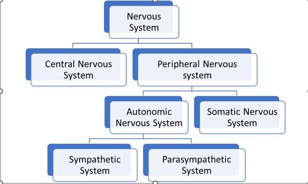
The human nervous system is divided into the somatic nervous system and the autonomic nervous system, each serving distinct functions:
Source of Sensory Information:
The somatic nervous system is primarily concerned with the external environment. It gathers sensory information from the skin, skeletal muscles, and joints.
The autonomic nervous system, on the other hand, monitors internal bodily functions. It receives input from visceral organs such as the heart, lungs, and digestive system.
Location of Receptors:
In the somatic system, sensory receptors are located on the body surface and within the musculoskeletal system.
In the ANS, receptors are found within internal organs and tissues, such as the stomach, lungs, and blood vessels.
Afferent Pathways:
Sensory neurons (afferents) in the somatic system enter the spinal cord through the dorsal root and typically end in the dorsal horn of the spinal cord.
In the ANS, afferent neurons also enter via the dorsal root but terminate in the intermediolateral horn (ILH) of the spinal cord, a region specialized for autonomic control.
Efferent Pathways:
The somatic nervous system uses a single motor neuron to connect the central nervous system (CNS) to its target: skeletal muscle.
The autonomic nervous system utilizes a two-neuron chain:
The preganglionic neuron originates in the brainstem or spinal cord (specifically in the ILH).
The postganglionic neuron resides in autonomic ganglia and extends to the target effector organ (such as smooth muscle or glands).
Effectors:
Somatic neurons innervate skeletal muscles, which are under voluntary control.
Autonomic neurons innervate smooth muscles, cardiac muscle, and glands, which are involuntary effectors.
Divisions of the ANS: Sympathetic vs. Parasympathetic
The ANS is divided into two primary branches, each with opposing actions:
| Feature | Sympathetic Nervous System | Parasympathetic Nervous System |
|---|---|---|
| Origin | Thoracolumbar outflow (T1–L2 spinal segments) | Craniosacral outflow (brainstem and sacral spinal segments S2–S4) |
| Preganglionic Fiber | Short, cholinergic (acts on nicotinic receptors) | Long, cholinergic (acts on nicotinic receptors) |
| Ganglia Location | Close to the spinal cord | Close to or within target organs |
| Postganglionic Fiber | Long, unmyelinated, usually adrenergic | Short, often myelinated, cholinergic |
| Postganglionic Neurotransmitter | Norepinephrine (acts on alpha- and beta-adrenergic receptors) | Acetylcholine (acts on muscarinic receptors) |
| Pre/Postganglionic Ratio | 1:100 (amplifies response) | 1:1 to 1:15 (localized response) |
| General Response | Widespread (“fight or flight”) | Targeted (“rest and digest”) |
| Energy Utilization | Mobilizes energy | Conserves energy |
Functional Impact on Organs
Here’s how each division affects various systems:
| Organ/System | Sympathetic Response | Parasympathetic Response |
|---|---|---|
| Eyes | Pupils dilate (mydriasis) | Pupils constrict (miosis) |
| Salivary Glands | Decreased secretion | Increased secretion |
| Sweat Glands | Increased sweating | Decreased sweating |
| Heart | Increased rate, conduction, and contraction | Decreased rate and contraction |
| Lungs | Bronchodilation | Bronchoconstriction |
| GI Tract | Decreased peristalsis, inhibited secretion | Increased peristalsis and secretion |
| Liver | Glycogen breakdown (glycogenolysis) | Glycogen synthesis (glycogenesis) |
| Urinary Bladder | Relaxes detrusor, contracts sphincter | Contracts detrusor, relaxes sphincter |
| Reproductive Organs (Male) | Ejaculation | Erection |
| Skin | Piloerection (goosebumps) | Relaxation of piloerector muscles |
Neurotransmitters and Receptors in the ANS
Autonomic signaling relies heavily on chemical messengers, or neurotransmitters, and their associated receptors. Understanding these is crucial for grasping how drugs and various stimuli affect autonomic function.
Cholinergic System (Acetylcholine-mediated)
Preganglionic neurons in both sympathetic and parasympathetic divisions release acetylcholine (ACh).
Postganglionic parasympathetic neurons also release ACh.
This system is referred to as cholinergic, and it acts on two primary types of receptors:
1. Nicotinic Receptors
Found at autonomic ganglia, the adrenal medulla, and the neuromuscular junction.
Activated by nicotine and ACh.
Mediate fast synaptic transmission via ligand-gated ion channels.
2. Muscarinic Receptors
Found in the heart, smooth muscles, and glands.
Activated by muscarine and ACh.
Have varied effects depending on the tissue:
Inhibitory in the heart (e.g., reduced heart rate),
Excitatory in the gastrointestinal system (e.g., increased motility and secretion).
Adrenergic System (Norepinephrine-mediated)
Postganglionic sympathetic neurons primarily release norepinephrine (noradrenaline).
This neurotransmitter acts on adrenergic receptors, which are classified as:
Alpha (α) receptors: Typically cause vasoconstriction and increased blood pressure.
Beta (β) receptors: Promote cardiac stimulation (β1) and bronchodilation (β2).
The adrenal medulla, considered a modified sympathetic ganglion, releases epinephrine (adrenaline) into the bloodstream, enhancing the sympathetic response.
Conclusion
The Autonomic Nervous System is a marvel of biological engineering, seamlessly managing the involuntary activities that keep us alive and responsive to our environment. Its two branches—sympathetic and parasympathetic—maintain a delicate balance between arousal and relaxation, ensuring that the body is always primed for either action or recovery. Understanding how these systems operate not only illuminates the intricacies of human physiology but also forms the foundation for many medical treatments targeting cardiovascular, respiratory, digestive, and neuropsychiatric disorders.
As we continue to explore the ANS, its influence on emotional health, stress regulation, and even immune responses becomes increasingly evident. This complex network truly exemplifies the body’s incredible ability to adapt, protect, and sustain itself—often without us even knowing it.