Classification, Habitat, Distribution, Life cycle, Laboratory Diagnosis, Treatment and Control of Ascaris lumbricoides
- Phylum – Nemathelminthes
- Class – Nematoda
- Genus – Ascaris
- Species – A. lumbricoides
The parasites are commonly referred as ‘Roundworms’. They are also called as Soil- Transmitted Helmiths (STH). A. lumbricoides are the largest human intestinal parasite, whose adult size ranges from (25-40)cm in length in female and (15-30)cm in male. Humans can also be infected by pig roundworm (Ascaris suum). Ascaris lumbricoides (human roundworm) and Ascaris suum (pig roundworm) are indistinguishable. Ascariasis is caused by ingesting eggs. This can happen when hands or fingers that have contaminated dirt on them are put in the mouth, or by consuming vegetables or fruits that have not been carefully cooked, washed, or peeled.
Habitat and geographical distribution of Ascaris
These parasites reside in human small intestine, specifically the jejunam. The geographic distributions of Ascaris lumbricoides are worldwide in areas with warm, moist climates. Infection occurs worldwide and is most common in tropical and subtropical areas where sanitation and hygiene are poor.
Life cycle of A. lumbricoides
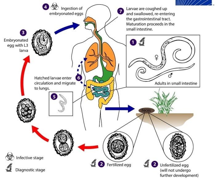
Adult worms live in the lumen of the small intestine. A female may produce approximately 200,000 eggs per day, which are passed with the feces. Unfertilized eggs may be ingested but are not infective. Larvae develop to infective fertile eggs after 18 days to several weeks, depending on the environmental conditions (optimum: moist, warm, shaded soil). After infective eggs are swallowed, the larvae hatch, invade the intestinal mucosa, and are carried via the portal, then systemic circulation to the lungs. The larvae mature further in the lungs (10 to 14 days), penetrate the alveolar walls, ascend the bronchial tree to the throat, and are swallowed. Upon reaching the small intestine, they develop into adult worms. Between 2 and 3 months are required from ingestion of the infective eggs to oviposition (egg production) by the adult female. Adult worms can live 1 to 2 years.

Clinical manifestation of Ascariasis
People infected with Ascaris often show no symptoms, regardless of the species of worm.
If symptoms do occur they can be light and include abdominal discomfort.
Heavy infections can cause intestinal blockage and impair growth/malnutrition in children.
Other symptoms such as cough are due to migration of the worms through the body. As larval stages travel through the body, they may cause visceral damage, peritonitis and inflammation, enlargement of the liver or spleen, and an inflammation of the lungs. Pulmonary manifestations take place during larval migration and may present as Loeffler’s syndrome, a transient respiratory illness associated with blood eosinophilia and pulmonary infiltrate with radiographic shadowing.
Laboratory diagnosis
i. Microscopy – The standard method for diagnosing ascariasis is by identifying Ascaris eggs in a stool sample using a microscope. Because eggs may be difficult to find in light infections, a concentration procedure is recommended.
ii. serology – During pulmonary disease, larvae may be found in fluids aspirated from the lungs. White blood cells counts may demonstrate peripheral eosinophilia.
Treatment, prevention and control
- albendazole and mebendazole, are the drugs of choice for treatment of Ascaris infections
- use of properly functioning and clean toilets by all community members as one important aspect; proper hand washing.
- Eliminating the use of untreated human faeces as fertilizer is also important
- in areas where more than 20% of the population is affected treating everyone is recommended. This has a cost of about 2 to 3 cents per person per treatment.This is known as mass drug administration and is often carried out among school-age children.