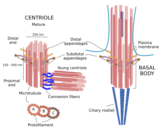
Centriole is a barrel shaped structure found in the area of centrosomes. Centrioles are physically made up of microtubules. The centrioles are in average 200nm in diameter and 500nm in length. Defect in centrioles are linked to cancer, microencephaly, blindness and are also said to be responsible in lung function.
Centrioles are the important subcellular structure with nine triplet microtubules in an animal cell that functions in cell-cell communication, cell division and cell motility. Centrioles are important in the differentiation of cells i.e. they direct asymmetrical divisions to drive cell differentiation, they are important for sensory function as well as lung function. They play a role in fertilization, however, the evidence to support this is still being studied. The ancestral and the conserved evolutionary function of centriole is to form cilia in the process of ciliogenesis. Though centrioles have not been found to contain DNA, yet they can form new centrioles with the help of massules which function as nucleating centers. The centrosome is a major organizer of the microtubular cytoskeleton in animal cells and helps in accurate cell division (mitosis and meiosis) and the pronuclei congregation (karyogamy) and govern cell architecture and polarization; for the formation of the centrosome, centriole is necessary. In animal cells, the centrioles facilitate the formation of the spindle fibers that separate the chromosomes into two halves during mitosis cell division This happens during the anaphase stage of mitosis in which the chromosomes of the cell move towards the different poles. Without centrioles, the chromosomes would not be able to move towards their poles.
Human sperm has centrioles, they are two remodeled centrioles which contributes in the zygote formation. Centrioles makes up the sperm aster and helps in the migration of pronuclei and the division. The centrioles in sperm are similar to that of other cells in early spermatogenesis but they get elongated as it reaches late spermatogenesis. The cell cycle of human centriole is similar to that of stem cell and lacks primary cilia in G1 phase. On contrast to sperm, oocyte lacks centrioles but contain PCM proteins which are dissolved in cytosol. After the sperm and oocyte fertilizes, the maternal PCM are recruited by the sperm centrioles to form zygote centrosomes.
Sperm centrosome remodeling takes place during spermiogenesis. During the remodeling process, there is loss and enrichment of centrosomal proteins and the formation of protein bars in DC. This DC develops to form an atypical centriole which has doublet microtubules instead of triplets. DC might enhance the sperm’s planar movement to the female reproductive tract. Remodeled DC will act to form centriole which participates in spindle pole formation. Providing a signal for differential fates of the zygote’s daughter cell is one of the potential roles of the atypical centriole. Atypical centriole may be important for sperm motility and early embryonic development. These findings now provide novel avenues for diagnostics and therapeutic strategies for male infertility and insights into early embryo developmental defects. These discoveries now provide innovative paths for diagnostics and therapeutic strategies for male infertility, and intuitions into early embryo developmental defects.
References Avidor-Reiss, T., & Fishman, E. (2019). It takes two (centrioles) to tango. Reproduction, 157(2), R33-R51. doi: 10.1530/rep-18-0350