The immune system is a highly intricate network of cells, tissues, and organs designed to safeguard the body from harmful pathogens and maintain overall health. Among its key components are lymph nodes, mucosa-associated lymphoid tissue (MALT), and cutaneous-associated lymphoid tissue (CALT), each playing a critical role in immune surveillance and response. Lymph nodes function as filtration points where lymphocytes interact with foreign antigens, while MALT and CALT serve as the body’s frontline defenses, monitoring mucosal surfaces and skin for potential threats. This guide provides an in-depth look at these components, their functions, and their roles in both innate and adaptive immunity, offering a thorough understanding of how the body mounts effective defenses against infections and diseases.
1. Lymph Nodes
1.1. Overview of Lymph Nodes
Lymph nodes are essential components of the lymphatic system, acting as filtration points for lymph fluid and serving as hubs for immune responses. They are scattered throughout the body and are pivotal in the body’s defense against pathogens.
- Function of Lymph Nodes: Lymph nodes filter lymph, a fluid that circulates through the lymphatic system, trapping foreign particles and pathogens. They provide a site for lymphocyte activation and proliferation.
1.2. Entry of Lymphocytes and Antigens
- Afferent Lymphatic Vessels: Lymphocytes and foreign antigens enter lymph nodes through afferent lymphatic vessels. This entry allows for the monitoring and initiation of immune responses within the node.
1.3. Lymphocyte Distribution in Lymph Nodes
- B Lymphocytes: These cells are primarily located in the follicles within the cortex of the lymph node. The cortex is the outer layer of the node and contains organized areas called follicles, where B cells encounter antigens. Upon activation, B cells differentiate into plasma cells that produce antibodies specific to the antigens.
- T Lymphocytes: T cells are predominantly found in the paracortex, which is the region between the cortex and the medulla. The paracortex is crucial for T cell activation, where they interact with antigen-presenting cells and become activated to carry out their immune functions.
2. MALT and CALT
2.1. Mucosa-Associated Lymphoid Tissue (MALT)
MALT is a critical component of the immune system located at mucosal surfaces. These surfaces are major entry points for pathogens and antigens.
- Locations and Function: MALT is found in mucosal areas of the gastrointestinal, respiratory, and urogenital tracts. It serves as a first line of defense against pathogens entering the body through these sites.
- Components of MALT: MALT is rich in macrophages and lymphocytes, which are essential for detecting and responding to antigens. Examples of MALT include:
- Tonsils: Located in the throat and mouth, these are among the first sites to encounter ingested or inhaled pathogens.
- Appendix: Positioned at the junction of the small and large intestines, the appendix contributes to the immune defense in the gut.
- Peyer’s Patches: Found in the small intestine, these lymphoid follicles monitor intestinal flora and pathogens.
2.2. Cutaneous-Associated Lymphoid Tissue (CALT)
CALT refers to the lymphoid tissue associated with the skin, providing an immune surveillance mechanism at the body’s largest organ.
- Components and Function: CALT contains various immune cells, including T cells, monocytes, macrophages, and dendritic cells, which are dispersed within the skin. These cells play a role in detecting and responding to antigens that come into contact with the skin.
3. Summary of Immune System Function
3.1. Innate Immunity
- Definition and Mechanism: Innate immunity is the body’s initial defense mechanism against infections, characterized by its nonspecific and immediate response. It includes physical barriers, such as the skin and mucous membranes, as well as various immune cells that act quickly upon encountering pathogens.
- Components:
- Neutrophils: Phagocytic cells that are among the first to respond to infection.
- Monocytes: These cells can differentiate into macrophages and dendritic cells, contributing to both innate immune responses and antigen presentation.
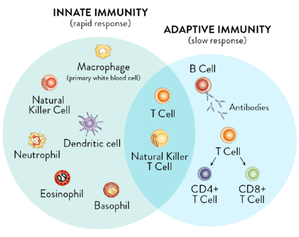
3.2. Adaptive Immunity
- Characteristics: Adaptive immunity is specific and involves a targeted response to particular pathogens. It is characterized by its ability to remember previous infections, providing a more effective response upon subsequent exposures.
- Key Components:
- B Cells: Develop in the bone marrow and, upon encountering antigens, differentiate into plasma cells that secrete specific antibodies.
- T Lymphocytes: Include several subtypes:
- Helper T Cells (Th): Assist other immune cells in their functions.
- Cytotoxic T Cells (Tc): Directly kill infected or cancerous cells.
- Regulatory T Cells (Treg): Regulate immune responses to maintain tolerance and prevent autoimmunity.
- Natural Killer (NK) Cells: Part of the innate immune system, NK cells can kill infected or cancerous cells without prior sensitization, providing an early defense mechanism.
3.3. Hematopoiesis and Blood Cells
- Hematopoietic Stem Cells: All blood cells, including those involved in immunity, arise from hematopoietic stem cells located in the bone marrow. These stem cells differentiate into various types of blood cells.
- Types of Peripheral Blood Leukocytes:
- Neutrophils: Key for early-stage immune responses and phagocytosis.
- Eosinophils: Combat parasitic infections and modulate allergic reactions.
- Basophils: Release histamines and other mediators involved in inflammatory responses.
- Monocytes: Transform into macrophages and dendritic cells in tissues, playing roles in both innate and adaptive immunity.
- Lymphocytes: Include B cells and T cells, which are crucial for the adaptive immune response.
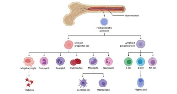
3.4. Tissue Cells Involved in Immunity
- Mast Cells: Found in tissues, particularly near blood vessels, they release histamines and other inflammatory mediators.
- Dendritic Cells: Act as antigen-presenting cells, capturing and presenting antigens to T lymphocytes.
- Macrophages: Phagocytic cells that engulf and digest pathogens and dead cells.
3.5. Lymphocyte Development and Function
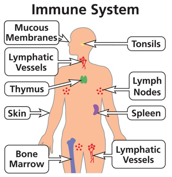
- Primary Lymphoid Organs:
- Bone Marrow: Site of B cell development and overall blood cell production.
- Thymus: Site of T cell maturation, where T cells gain the ability to recognize specific antigens.
- Secondary Lymphoid Organs:
- Lymph Nodes: Sites where mature lymphocytes encounter antigens and become activated.
- Spleen: Filters blood and supports lymphocyte activation and proliferation.
- MALT and CALT: Sites of immune surveillance and response at mucosal surfaces and the skin, respectively.
This detailed summary provides an overview of lymph nodes, MALT, CALT, and the immune system’s innate and adaptive components. It is essential for understanding how the body defends itself against pathogens and maintains overall health.