Author : Roshni Nepal Phylum – Nemathelminthes
Class – Nematoda
Genus – Ancylostoma, Necator
Species – A. duodenale, N. americanus
Intestinal hookworm disease in humans is caused by Ancylostoma duodenale, Necator americanus. They are also called as Soil- Transmitted Helmiths (STH). Hookworm eggs are passed in the feces of an infected person. Hookworm infection is mainly acquired by walking barefoot on contaminated soil. Filariform (L3) larvae are the infective form. On contact with the human host, typically bare feet, the larvae penetrate the skin.
Habitat and geographical distribution
The geographic distributions of the hookworm species that are intestinal parasites in human, Ancylostoma duodenale and Necator americanus, are worldwide in areas with warm, moist climates. Hookworms live in the small intestine.
Life cycle of intestinal hookworm parasites

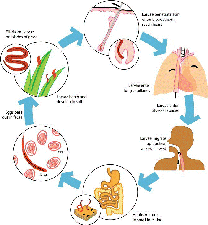
Eggs are passed in the stool, and under favorable conditions (moisture, warmth, shade), larvae hatch in 1 to 2 days and become free-living in contaminated soil. These released rhabditiform larvae grow in the feces and/or the soil, and after 5 to 10 days (and two molts) they become filariform (third-stage) larvae that are infective. These infective larvae can survive 3 to 4 weeks in favorable environmental conditions. On contact with the human host, typically bare feet, the larvae penetrate the skin and are carried through the blood vessels to the heart and then to the lungs. They penetrate into the pulmonary alveoli, ascend the bronchial tree to the pharynx, and are swallowed. The larvae reach the jejunum of the small intestine, where they reside and mature into adults. Adult worms live in the lumen of the small intestine, typically the distal jejunum, where they attach to the intestinal wall with resultant blood loss by the host. Most adult worms are eliminated in 1 to 2 years, but the longevity may reach several years.
Some A. duodenale larvae, following penetration of the host skin, can become dormant but are capable of re-activating and establishing patent, intestinal infections.
Clinical manifestation of hookworms
High-intensity hookworm infections occur among both school-age children and adults The most serious effects of hookworm infection are the development of anemia and protein deficiency caused by blood loss at the site of the intestinal attachment of the adult worms.
When children are continuously infected by many worms, the loss of iron and protein can retard growth and mental development.
Laboratory diagnosis
The standard method for diagnosing the presence of hookworm is by identifying hookworm eggs in a stool sample using a microscope. Because eggs may be difficult to find in light infections, a concentration procedure is used.
The eggs are oval or elliptical, measuring 60 by 40 µm, colorless, non bile stained and with a thin transparent hyaline shell membrane.
Treatment, prevention and control
- albendazole and mebendazole, are the drugs of choice for treatment of hookworm infections. Infections are generally treated for 1-3 days; iron supplements are prescribed for anaemic patients.
- The best way to avoid hookworm infection is not to walk barefoot in areas where hookworm is common and where there may be human fecal contamination of the soil. Also, avoid other skin contact with such soil and avoid ingesting it.
- Infection can also be prevented by not defecating outdoors and by effective sewage disposal systems.