- Apoptosis is a physiological process of programmed cell death and occurs in multicellular organisms. The word ‘apoptosis’ is derived from a Greek word that means ‘falling off’.
- When the cells are no longer required for the body, it automatically commits suicide by activating an intracellular death program. Therefore, it is called a programmed cell death.
- In a biological mechanism, it is a cell that undergoes self-destruction when stimulated by the mild cellular injuries and by external or internal factors of the cell.
- Normally, apoptosis occurs during the development and aging of the body’s tissue and cells to maintain the cell population.
- Mainly biochemical factors like cell shrinkage, nuclear fragmentation, nucleus and cytoplasm condensation, chromosomal DNA fragmentation, and decaying of mRNA lead to change in morphology of the cells and cause death.
- Death of cells actually balances the cell division or else there will be unwanted growth of cells that leads to cancer and development of tumors.
- The average adult somewhat loses 50 to 70 billion cells per day due to apoptosis. Too little and too much cell death leads to many diseases.
- Apoptosis and necrosis both cause cell death but in different ways. Necrosis is the process of cell death that harms them with toxic chemicals or physical injuries and causes inflammation whereas apoptosis is a well-programmed death of cells.
Morphological and biochemical changes during apoptosis:

(Source: https://www.quora.com/How-does-apoptosis-occur-1)
- Apoptosis of cells undergoes an orderly process and requires several hours for the initiation of the final stage of cell death.
- The time taken depends according to the cell type and the apoptotic pathway that it undergoes.
- In the initial stage of cell death, the cell shrinks and develops blebs on the surface which are bubble-like protrusions. The DNA and endoplasmic reticulum gets fragmented into small pieces. Similarly, pyknosis and retraction of pseudopods occur where the entire cell splits up in the end.
- The split small chunks of the cell are neatly packed in a membrane and start releasing signals attracting phagocytic cells. In the case of dying cells, they produce phosphatidylserine which is a lipid molecule that attracts phagocyte.
- During apoptosis, three main types of biochemical changes occur. Caspases get activated, breakdown of DNA and proteins and phagocytic cell recognizes during membrane change.
Mechanisms of apoptosis:
The mechanism of apoptosis is highly complex that it requires energy cascade and also helps in the understanding of pathogenesis.
There are two main apoptotic pathways where caspases are central to both the mechanism. Below are the two commonly described pathways:
Intrinsic pathway (or mitochondrial pathway) and the extrinsic pathway (or death receptor pathway).
(Source:https://teachmephysiology.com/biochemistry/cell-growth-death/apoptosis/)
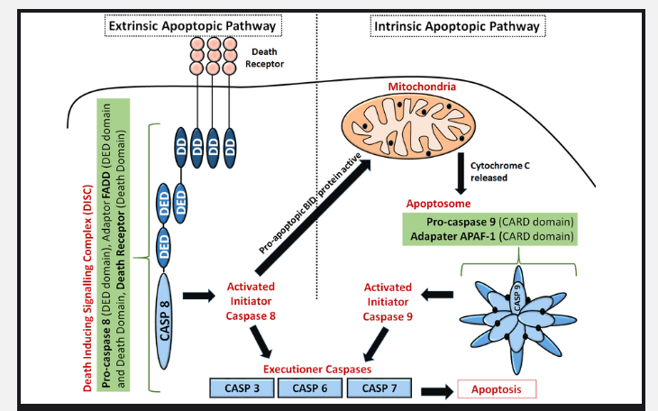
Intrinsic (or mitochondrial) pathway:
- The intrinsic pathway is known to be the mitochondrial pathway because the apoptotic proteins target the mitochondrial cell in various ways like forming membrane pores that cause swelling of mitochondria or increasing permeability and causing leakage.
- To initiate the pathway, some internal stimuli must be present like irreparable genetic damage, hypoxia, high concentration of cytosolic Ca2+, and oxidative stress.
- These stimuli trigger the pathway and increase mitochondrial permeability where cytochrome-c molecules release out into the cytoplasm.
- This occurs through the action of Bax and Bak proteins. The cytochrome- c then binds with the Apoptotic protease activating factor-1 (Apaf-1) and ATP which again binds to procaspase 9 and forms a complex protein, apoptosome.
- This then activates caspase 3 while on the other side, SMACs (second mitochondria-derived activator of caspases) or Omi/HtrA2 binds to the inhibitor of apoptosis proteins (IAPs) and promotes caspase activation.
- A molecular switch in response to the stimuli determines the cell to survive or undergo apoptosis.
Extrinsic (or death receptor) pathway:
- External stimuli trigger the apoptosis by binding specific ligands at the death receptor on the cell surface.
- Type 1 TNF receptor (TNFR1) is the best-known death receptor and a related protein Fas (CD95) with their ligands TNF and Fas ligand.
- These two TNF induced model and the Fas-Fas ligand-mediated model are the two ways of initiation of apoptotic mechanism in mammals where both involve TNF receptors.
- TNF induced model is also known as tumor necrosis factor and its TNF-alpha is the major extrinsic mediator. The TNF-alpha is activated by the macrophages which bind to the TNFR1 and initiate caspase activation. The abnormal production of TNF-alpha leads to several autoimmune diseases.
- Fas-Fas ligand-mediated model is the interaction between Fas and FasL and forms a death-inducing signaling complex (DISC). DISC contains the FADD, caspase 8, and 10 which directly activates other caspase families and triggers the cell apoptosis.
The common pathway
The execution phase involves the activation of a number of caspases. The intrinsic pathway activates caspase 9 while the extrinsic pathway activates caspase 8 then both converge to caspase 3 which cleaves the inhibitor of the caspase-activated deoxyribonuclease. Downstream caspases induce protein kinase, DNA repair protein, and cytoskeleton protein cleavage and also effects cell cycle and signaling pathways.
References:
- https://www.khanacademy.org/science/biology/developmental-biology/apoptosis-in-development/a/apoptosis#:~:text=Apoptosis%20is%20an%20orderly%20process,maintains%20balance%20in%20the%20body.
- https://en.wikipedia.org/wiki/Apoptosis
- https://www.ncbi.nlm.nih.gov/pmc/articles/PMC2117903/
- https://www.ncbi.nlm.nih.gov/books/NBK26873/
- https://www.genome.gov/genetics-glossary/apoptosis
- https://jeccr.biomedcentral.com/articles/10.1186/1756-9966-30-87
- https://www.medicinenet.com/script/main/art.asp?articlekey=11287
- https://www.britannica.com/science/apoptosis
- https://teachmephysiology.com/biochemistry/cell-growth-death/apoptosis/