- Gram-positive bacteria retain the crystal violet dye by its thick layer of peptidoglycan and appear purple color when observed under the microscope after performing Gram staining.
- Crystal violet is a water-soluble dye that enters the peptidoglycan layer and forms crystal violet-iodine complexes.
- Then, the 95% ethyl alcohol acts as a decolorizer that dehydrates and shrinks the peptidoglycan layer making the formed complexes unable to escape that result in purple stained cells.
- Due to its low lipid and lipoprotein content in its cell wall, gram-positive bacteria are highly susceptible to antibiotics but are resistant to physical disruption because of its multilayered peptidoglycan.
- Gram staining is the first step for the identification of an unknown bacterial culture followed by specific biochemical tests as described in Bergey’s manual.
- Morphological observation of culture colonies is also observed before these tests.
Discover gram positive bacteria with accuracy using biochemical tests for identification . Learn more in this article.
Morphological test
The colony morphological characteristics are observed from the culture plate. Shape, size (in mm), color, consistency, opacity, elevation, and margin are determined and noted down for further identification.
Gram staining for identification of gram positive bacteria
Gram staining is a differential staining technique that differentiates bacteria on its ability to retain color. Gram-positive bacteria retain purple color from the primary dye while gram positive bacteria retain the counterstain color and appear pink or red color when visualized under a microscope.
The cell wall composition of gram-negative and gram-positive bacteria differs that is why the mechanism of Gram staining in the decolourizing step acts differently in both of the bacteria resulting in two different colors for identification.
Some of the specific biochemical tests for the identification of gram positive bacterial species are:
- Catalase test
- Oxidative/Fermentative
- Bacitracin susceptibility test
- Bile Esculin test
- Hippurate hydrolysis test
- Coagulase test
Catalase test
- The catalase test determines the organism’s ability to produce catalase enzyme which forms gas bubbles when reacting with 3% H2O2.
- Catalase producing gram-positive organisms:
S. aureus, Staphylococcus spp, Micrococcus spp.
Oxidative/Fermentative Test
- During the metabolism process, bacteria either break down complex organic molecules aerobically or anaerobically.
- The aerobic metabolism process is referred to as the oxidation process whereas the anaerobic metabolism process is referred to as a fermentative process.
- Two tubes with Hugh and Leifson’s medium are used where one tube is sealed with paraffin oil to create an anaerobic condition.
- Oxidative gram-positive organism:
Micrococcus spp.
- Fermentative gram positive organism:
S. aureus
Bacitracin susceptibility test
- Bacitracin is antibiotic produced by the licheniformis group of Bacillus subtilis var Tracy which disrupts the cell wall of the gram-positive bacteria.
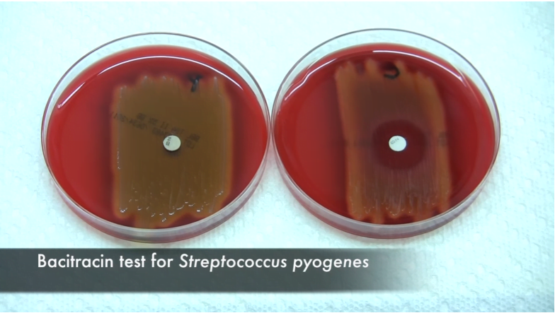
- Bacitracin susceptibility test is a presumptive test for the differentiation between beta-hemolytic Group A streptococci and beta-hemolytic non-Group A streptococci.
- This test is performed on the blood agar with a streaked culture of streptococci where the bacitracin disk is impregnated with sterile forceps before incubation.
- A zone of inhibition will be observed if the isolated organism is beta-hemolytic Group A streptococci whereas beta-hemolytic non-Group A streptococci will be resistant towards bacitracin showing no zone of hydrolysis and will grow all over the disk.
- Bacitracin sensitive Group A streptococci:
S. pyogens
- Bacitracin resistant non-Group A streptococci:
S. agalactiae

Bile Esculin test
- Bile esculin agar medium is both selective as well as the differential medium in which its selective ingredient is bile that inhibits the growth of other gram-positive bacteria except enterococci and some streptococci species.
- Esculin is the differential ingredient that differentiates Enterococcus from Streptococcus.
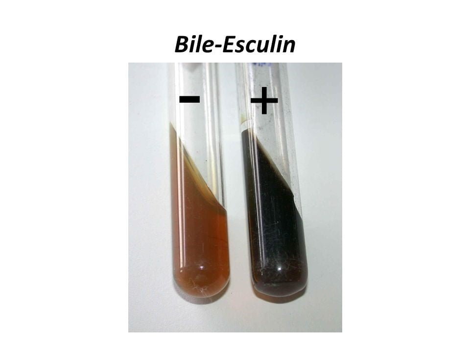
- The bile esculin test determines the ability of organisms to hydrolyze esculin when in the presence of bile, esculetin is formed.
- Ferric citrate is present in the medium and when it reacts with esculetin, it turns the entire medium dark brown to black due to the formation of the phenolic iron complex.
- Bile esculin gram-positive organism:
Enterococcus faecalis

Hippurate hydrolysis test
- The Hippurate hydrolysis test detects the organism’s ability to produce hippuricase enzyme that hydrolyzes hippurate substrate into glycine and benzoic acid.
- Glycine is detected by using the Ninhydrin reagent which forms a deep purple or violet color.
- The Hippurate hydrolysis test differentiates β-hemolytic Streptococcus agalactiae from other β-hemolytic streptococci.
- Hippurate gram-positive organism:
Streptococcus agalactiae

Coagulase test
- The coagulase test determines the organism producing the coagulase enzyme which is Staphylococcus aureus.
- This test helps in the differentiation between coagulase-positive Staphylococcus aureus and coagulase-negative Staphylococcus (CONS).
- S. aureus produces a bound and free form of coagulase that converts soluble fibrinogen into insoluble fibrin.
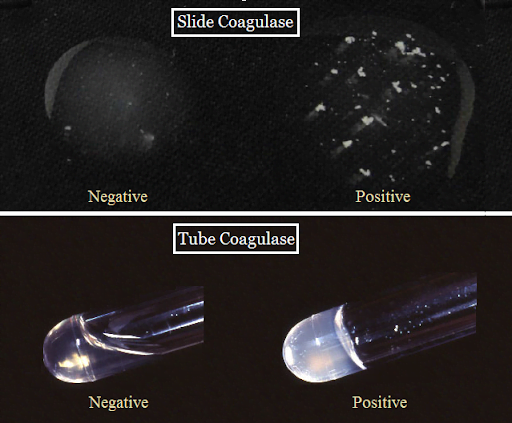
- Coagulase test is done either in a slide or in a tube which is determined by the form of coagulase produced.
- Cell bound coagulase is detected by the slide coagulase test which forms agglutination in case of positive results.
- Free coagulase is detected in a tube which forms a clot if the organism is tested positive.
- Coagulase producing gram-positive organism:
Staphylococcus aureus.

(Source: http://laboratorytests.org/coagulase-test/ )
References:
- https://www.technologynetworks.com/immunology/articles/gram-positive-vs-gram-negative-323007
- http://shs-manual.ucsc.edu/policy/bacitracin-test-disk
- http://www.uwyo.edu/molb2021/additional_info/summ_biochem/bile_esculin.html#:~:text=This%20is%20a%20medium%20that,Enterococcus%20(E%20faecalis%20and%20E.
- Manandhar.S,Sharma.S,(2013).Practical Approach to Microbiology,National Book Center,2nd edition (Pageno.65-81).