Ribosomes are cellular structures responsible for protein synthesis. Composed of RNA and protein, they decode mRNA sequences, add amino acids, and play a crucial role in protein production in cells.
What are Ribosomes?
- Ribosomes, also known as ribonucleoproteins, are intercellular structures responsible for protein synthesis in cells.
- They function as complex molecular machines found in both prokaryotic and eukaryotic cells.
- Ribosomes are composed of RNA and protein components.
- They are not membrane-bound and can be located in different parts of the cell, such as the cytosol, rough endoplasmic reticulum, or specific locations.
- During protein synthesis or translation, ribosomes read the sequence of messenger RNA (mRNA) and translate it into a specific sequence of amino acids.
- Ribosomes bind to mRNA and decode the information encoded in its nucleotide sequence.
- They interact with transfer RNAs (tRNAs) carrying amino acids, adding them to the growing protein chain.
- The process of protein synthesis involves peptidyl transfer, elongation, and fidelity of translation.
- Ribosomal RNA (rRNA) molecules and ribosomal proteins facilitate these steps.
- Specific factor proteins catalyze different stages of protein synthesis.
- Ribosomes are specialized organelles that serve as sites of protein synthesis.
- They play a vital role in the production of functional proteins in all living cells.
Composition and Structure of Ribosomes
Composition of Ribosomes
- Ribosomes are made up of ribosomal RNA (rRNA) and proteins.
- In prokaryotic cells, there are three types of rRNA: 16S rRNA, 23S rRNA, and 5S rRNA.
Subunits and Densities
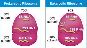
- Ribosomes consist of two subunits with densities of 50S and 30S.
- The 30S subunit contains 16S rRNA and 21 proteins, while the 50S subunit contains 5S and 23S rRNA and 31 proteins.
- During protein synthesis, these subunits combine to form a complete 70S ribosome.
Localization and Structure of Ribosomes
- Ribosomes are located in the cytosol of cells, either scattered throughout or attached to the endoplasmic reticulum.
- They have two areas of cytoplasmic localization.
- The structure of free and bound ribosomes is similar, and they are involved in protein synthesis.
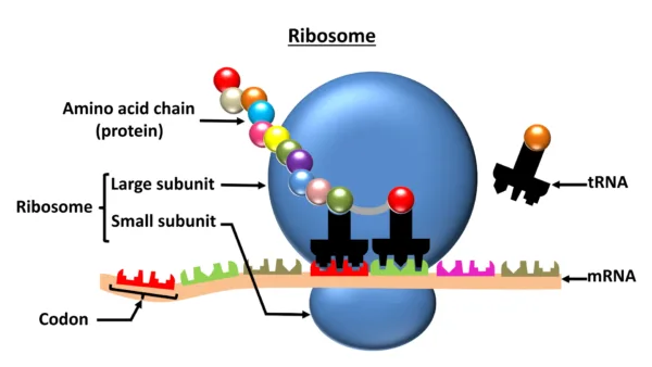
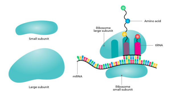
Function and Structure of Ribosomes
- Each ribosome consists of a smaller subunit and a larger subunit.
- The smaller subunit binds to mRNA and decodes the genetic message, while the larger subunit catalyzes peptide bond formation and binds to aminoacylated tRNAs.
- Both subunits contain ribonucleic acid (RNA) and protein components.
Binding Sites and tRNA Interaction
- Ribosomes have three binding sites for tRNA: A site, P site, and E site.
- The A site acknowledges the approaching aminoacylated tRNA, the P site holds the tRNA with the growing peptide chain, and the E site holds the deacylated tRNA before it leaves the ribosome.
Svedberg Units and Ribosomal Densities
- The Svedberg (S) units describe the density of ribosomal subunits during centrifugation.
- Prokaryotic cells have 70S ribosomes, while eukaryotic cells have 80S ribosomes.
- Eukaryotic ribosomes consist of 40S and 60S subunits, while prokaryotic ribosomes have 30S and 50S subunits.
- The Svedberg values are not additive, as sedimentation rate depends on size and shape, not just molecular weight.
Ribosomes, composed of rRNA and proteins, play a crucial role in protein synthesis. Prokaryotic cells have 30S and 50S subunits, forming 70S ribosomes. Ribosomes are localized in the cytosol, either scattered or attached to the endoplasmic reticulum. They consist of smaller and larger subunits, each with specific functions in mRNA decoding and peptide bond formation. Ribosomes have binding sites for tRNA and are characterized by their Svedberg densities. Understanding the composition and structure of ribosomes provides insights into their fundamental role in cellular processes.
Prokaryotic Ribosomes: Composition and Structure
- Prokaryotic ribosomes are made up of two subunits: the 30S subunit and the 50S subunit.
- These subunits combine to form a complete ribosome with a density of 70S.
Function of the 30S Subunit
- The 30S subunit contains the 16S ribosomal RNA (rRNA) and 21 proteins.
- The 16S rRNA is essential for decoding genetic information carried by messenger RNA (mRNA) during translation.
- It interacts with mRNA, recognizes the start codon, and facilitates binding with the initiator transfer RNA (tRNA).
Role of the 50S Subunit
- The 50S subunit consists of two types of rRNA: the 23S rRNA and the 5S rRNA.
- The 23S rRNA catalyzes peptide bond formation during protein synthesis.
- The 5S rRNA provides structural stability to the ribosome.
Ribosomal Proteins and Structure
- Both subunits contain various ribosomal proteins that contribute to the overall structure and function of the ribosome.
- The specific number and composition of these proteins may vary slightly among different prokaryotic species.
Protein Synthesis and Ribosome Function
- The 30S and 50S subunits combine to form a complete 70S ribosome responsible for protein synthesis in prokaryotic cells.
- Ribosomes bind to mRNA and coordinate the binding of transfer RNAs (tRNAs) carrying amino acids.
- This enables the assembly of amino acids in the correct order based on the mRNA sequence.
Differences Between Prokaryotic and Eukaryotic Ribosomes
- Prokaryotic ribosomes are smaller compared to eukaryotic ribosomes, which have 80S ribosomes consisting of a 40S subunit and a 60S subunit.
- The variations in ribosomal subunits between prokaryotes and eukaryotes present targets for selective inhibition by antibiotics.
- Different organisms can have distinct structural and functional characteristics in their ribosomes.
Prokaryotic ribosomes are composed of the 30S and 50S subunits, forming a functional 70S ribosome for protein synthesis. These ribosomes contain rRNA, ribosomal proteins, and play crucial roles in mRNA decoding and peptide bond formation. Prokaryotic ribosomes differ in size and subunit composition from eukaryotic ribosomes, providing opportunities for targeted antibiotic inhibition. Overall, prokaryotic ribosomal subunits serve as essential components in protein synthesis processes.
Eukaryotic Ribosomes: Composition and Structure
- Eukaryotic ribosomes consist of two subunits: the 40S subunit and the 60S subunit.
- These subunits combine to form a complete ribosome with a density of 80S.
Function of the 40S Subunit
- The 40S subunit contains the 18S ribosomal RNA (rRNA) and approximately 33 ribosomal proteins.
- The 18S rRNA decodes mRNA and initiates translation by identifying the start codon.
Role of the 60S Subunit
- The 60S subunit comprises three types of rRNA: 5.8S rRNA, 28S rRNA, and 5S rRNA.
- The 5.8S and 28S rRNAs catalyze peptide bond formation and provide structural stability.
- The 5S rRNA contributes to the overall structure and function of the ribosome.
Additional Ribosomal Proteins
- Both subunits contain extra ribosomal proteins that maintain structural integrity and aid in translation.
Protein Synthesis and Ribosome Function
- The combination of the 40S and 60S subunits forms an 80S ribosome responsible for protein synthesis in eukaryotic cells.
- Ribosomes bind to mRNA and coordinate the binding of transfer RNAs (tRNAs) carrying amino acids.
- This facilitates the assembly of amino acids into a polypeptide chain following the mRNA sequence.
Differences between Eukaryotic and Prokaryotic Ribosomes
- Eukaryotic ribosomes are larger and more complex than prokaryotic ribosomes.
- Structural and functional variations between these ribosomes make them potential targets for antibiotics.
Ribosomes and the Endoplasmic Reticulum (ER)
- In eukaryotes, ribosomes are present not only in the cytoplasm but also associated with the endoplasmic reticulum (ER).
- ER-bound ribosomes play a role in synthesizing proteins for secretion or membrane insertion.
Eukaryotic ribosomes consist of the 40S and 60S subunits, which combine to form an 80S ribosome involved in protein synthesis. These ribosomes contain rRNA and ribosomal proteins that decode mRNA, catalyze peptide bond formation, and maintain structural integrity. Eukaryotic ribosomes are larger and more complex than prokaryotic ribosomes, providing potential targets for selective antibiotics. Additionally, ribosomes associated with the endoplasmic reticulum participate in synthesizing proteins for secretion or membrane insertion.
Functions of Ribosomes
The functions of ribosomes can be summarized as follows:
1. Protein Synthesis:
Ribosomes are responsible for protein synthesis, which is crucial for cell activities and the functioning of living organisms. They serve as the site where genetic instructions encoded in mRNA are translated into proteins. Ribosomes facilitate the assembly of amino acids into polypeptide chains according to the sequence of mRNA.
2. Transcription and Translation:
Ribosomes are involved in both transcription and translation processes. During transcription, DNA is transcribed into mRNA, which carries the genetic information from the nucleus to the cytoplasm where ribosomes are located. In the cytoplasm, ribosomes bind to the mRNA and facilitate the translation process, where the genetic code is decoded to synthesize proteins.
3. mRNA Decoding:
Ribosomes possess the ability to decode the mRNA code. The ribosome moves along the mRNA template and reads each codon (a sequence of three nucleotides) of the mRNA. It pairs each codon with a specific amino acid carried by a transfer RNA (tRNA), which has a complementary anticodon on one end and the corresponding amino acid on the other end. This process ensures that the correct amino acids are incorporated into the growing polypeptide chain.
4. Protein Folding:
Ribosomes also play a role in protein folding. As the polypeptide chain emerges from the ribosome, it undergoes folding to acquire its functional three-dimensional structure. Ribosomes, along with other cellular factors such as chaperone proteins, assist in the proper folding of proteins, ensuring their functionality.
5. Co-translational Processes:
Ribosomes are involved in co-translational processes that occur simultaneously with protein synthesis. These processes include co-translational modifications, such as glycosylation or phosphorylation, which can occur as the nascent polypeptide chain is being synthesized by the ribosome. Additionally, the targeting of proteins to specific cellular compartments or organelles often happens co-translationally, with ribosomes synthesizing proteins that contain specific signal sequences directing their localization.
6. Subcellular Localization:
Ribosomes can be found in different subcellular locations. In addition to free ribosomes in the cytoplasm, ribosomes can associate with the endoplasmic reticulum (ER) to form the rough ER. Ribosomes bound to the ER are involved in the synthesis of proteins that are destined for secretion, membrane insertion, or incorporation into the ER itself.
In summary, ribosomes are essential cellular organelles that facilitate protein synthesis, decode mRNA, aid in protein folding, and participate in various co-translational processes. Their functions are critical for the proper functioning of cells and the production of functional proteins necessary for life processes.
Ribosomopathies: Diseases Associated with Ribosome Dysfunction
Ribosomes are fundamental cellular structures involved in protein synthesis, playing a vital role in maintaining cellular function. However, when ribosome function is impaired, it can lead to a group of disorders known as ribosomopathies. This article explores the concept of ribosomopathies, their underlying causes, and the impact they have on human health.
What are Ribosomopathies?
Ribosomopathies are a collection of rare genetic disorders characterized by defects in ribosome function. These disorders can manifest in various ways, affecting multiple organ systems. Ribosomopathies can result from mutations in ribosomal proteins, ribosomal RNA, or factors involved in ribosome biogenesis.
Common Ribosomopathies
Several ribosomopathies have been identified, including Diamond-Blackfan anemia, Shwachman-Diamond syndrome, and 5q- syndrome. These conditions present with distinct clinical features, such as bone marrow failure, skeletal abnormalities, and increased susceptibility to certain cancers. Ribosomopathies can have diverse clinical manifestations due to the essential role of ribosomes in all cells.
Ribosome dysfunction disrupts protein synthesis, leading to cellular stress, impaired growth, and developmental abnormalities. In ribosomopathies, defective ribosomes can trigger cellular responses, including altered gene expression and apoptosis. These abnormalities contribute to the pathogenesis of ribosomopathies and the observed clinical phenotypes.
Understanding the molecular mechanisms underlying ribosomopathies is crucial for developing targeted therapies. Researchers are investigating potential therapeutic strategies, such as modulating ribosome biogenesis, restoring ribosome function, or targeting specific pathways affected by ribosome dysfunction. Currently, treatment mainly focuses on managing symptoms and complications associated with ribosomopathies.
References
- Alberts, B., Johnson, A., Lewis, J., Raff, M., Roberts, K., & Walter, P. (2002). Molecular Biology of the Cell. Garland Science.
- Lodish, H., Berk, A., Zipursky, S. L., Matsudaira, P., Baltimore, D., & Darnell, J. (2000). Molecular Cell Biology. W. H. Freeman.
- Nelson, D. L., Cox, M. M. (2008). Lehninger Principles of Biochemistry. W. H. Freeman.
- Ramakrishnan, V. (2002). Ribosome structure and the mechanism of translation. Cell, 108(4), 557-572.
- Ban, N., Beckmann, R., Cate, J. H., Dinman, J. D., Dragon, F., Ellis, S. R., … & Yonath, A. (2014). A new system for naming ribosomal proteins. Current Opinion in Structural Biology, 24, 165-169.
- Woolford, J. L., & Baserga, S. J. (2013). Ribosome biogenesis in the yeast Saccharomyces cerevisiae. Genetics, 195(3), 643-681.
- Henras, A. K., Soudet, J., Gérus, M., Lebaron, S., Caizergues-Ferrer, M., Mougin, A., & Henry, Y. (2008). The post-transcriptional steps of eukaryotic ribosome biogenesis. Cellular and Molecular Life Sciences, 65(15), 2334-2359.
- Ruggero, D., & Shimamura, A. (2014). Marrow failure: a window into ribosome biology. Blood, 124(18), 2784-2792.
- Mills, E. W., & Green, R. (2017). Ribosomopathies: there’s strength in numbers. Science, 358(6363), eaan2755.
- Narla, A., & Ebert, B. L. (2010). Ribosomopathies: human disorders of ribosome dysfunction. Blood, 115(16), 3196-3205.