- Meiosis is a form of eukaryotic cell division process where a single cell divides twice and produces four gamete cells that contains the reduced number of chromosomes than that in the parent cell.
- They produces haploid sex cells or gametes i.e, they contain half the number of each copy of chromosome from diploid cells that contains two copies of each chromosome.
- Meiosis cell division process describes a specific process of cell division by which gametes are made where egg and sperm cells are produced for sexual reproduction.
- When the sperm cell and egg cell fuse during reproduction and forms a single cell called zygote, the number of chromosomes restores in the offspring.
- Meiosis is important for many sexually reproducing animals to make sure that the offspring contains same number of chromosomes as in the parents.
- In many animals, if the number of alleles in each gene is not reduced to one in the gametes, there will be four copies of each gene in the offspring; this will lead to many developmental defects.
- In other organisms, they contain more than two sets of chromosomes which are known as polyploidy. It is a heritable condition that occurs in plants like peanuts, apples, and bananas, some reptiles, amphibians and insects are also seen to have polyploidy.
- However, if the organism cannot survive being polyploidy, they must undergo meiosis before reproduction.
Meiosis cell division process is somewhat similar to mitosis. Meiosis begins from the DNA replication in the male and female reproductive organs and the process splits into meiosis I and meiosis II where both divisions have multiple phases.
In meiosis I, the duplicated DNA separates into daughter cells while in meiosis II, the two alleles of each gene separates into individual cells.
Phases of Meiosis I:
- Interphase
- Prophase I
- Metaphase I
- Anaphase I
- Telophase I and cytokinesis
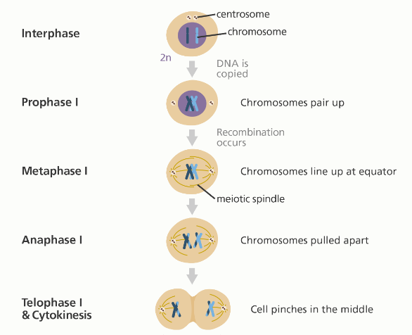
Interphase
- In this preliminary phase, the cell DNA is copied which results in two identical full sets of chromosomes.
- During interphase, outside of the nucleus contains two centrosomes where microtubules extend from.
- Each centromere contains a pair of centrioles.
Prophase I
- Each chromosome comprises of two sister chromatids condensing and forming X shaped structure.
- Both chromosome pairs up so that copies of chromososme I and II exchange nuclear materials by the recombination or crossing over process.
- The membrane around the nucleus starts to dissolve at the end of the prophase I and releases the chromosomes.
Metaphase I
- The released chromosome pairs up the line next to each other along the equator of the cell.
- The centriole remains at the opposite poles with the meiotic spindle fiber (consists of microtubules and other proteins) extending from them where each pair of chromosome attaches to it.
Anaphase I
- The meiotic spindle fiber pulls apart each pair of chromosome to one pole and to another pole of the cells.
- The sister chromatids stay together in this division which is different from mitosis and meiosis II.
Telophase I and cytokinesis:
- The chromosomes continue to move to the opposite poles of the cell until a full set of chromosomes gather together.
- In each set of chromosomes, a membrane is formed to create two new nuclei.
- Each single cell squeezes at the middle forming two daughter cells with a full set of chromosomes in each cell. This process is cytokinesis.
Phases of Meiosis II
- Prophase II
- Metaphase II
- Anaphase II
- Telophase II and cytokinesis
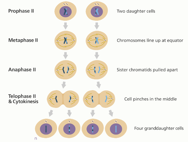
Prophase II:
- Two daughter cells formed in the meiosis I contains 23 pairs of chromatids each.
- The chromosomes starts to condense again and forms X shaped structure.
- The nuclear membrane starts to dissolve and releases chromosomes.
- The centrioles duplicate and meiotic spindle fiber forms again.
Metaphase II:
- The chromosomes of each daughter cell lines up along the equator and the centrioles remain at the two opposite pole along with each daughter cell.
- Meiotic spindle fiber gets attached to each of the sister chromatids.
Anaphase II:
- The meiotic spindle fiber pulls the sister chromatids to opposite poles and gets separated.
- The separated chromatids become individual chromosome.
Telophase II and cytokinesis:
- The separated chromatids continue to move towards the opposite pole of the cell until a full set of chromosomes gather together.
- In each set of chromosome, a membrane forms to form two new nuclei cell.
- Even though this is a last phase of meiosis cell division, cytokinesis completes the process.
- Cytokinesis process forms four granddaughter cells, each containing a haploid (half) set of chromosomes.
- In males, these four granddaughter cells are the sperm cells while in female, one is an egg cell and remaining three are polar bodies that do not develop into eggs.
References:
- https://www.nature.com/scitable/definition/meiosis-88/
- https://biologydictionary.net/meiosis/
- https://teachmephysiology.com/biochemistry/cell-growth-death/meiosis/
- https://www.khanacademy.org/science/ap-biology/heredity/meiosis-and-genetic-diversity/a/phases-of-meiosis
- https://www.yourgenome.org/facts/what-is-meiosis
- https://www2.le.ac.uk/projects/vgec/highereducation/topics/cellcycle-mitosis-meiosis