Our eyes serve as our body’s photoreceptors since they are the organ of vision. The human eye is a hollow, spherical organ that is 24 mm in diameter and weighs 6–8 gm. It is also known as an eyeball because of its form. The eye can gaze up, down, and side to side because of the
Six sets of muscles that extend from the eye orbit to the outer surface of the eyeball. There are two categories of muscles. i. Rectus muscles – superior rectus, internal rectus, inferior rectus and external rectus, ii. Oblique muscles – superior oblique and inferior oblique.
The eye ball consists of 3 concentric layers.:
- The outer fibrous layer
- The middle vascular layer
- Retina or inner layer
The outer fibrous layer: It is made up of 3 parts.
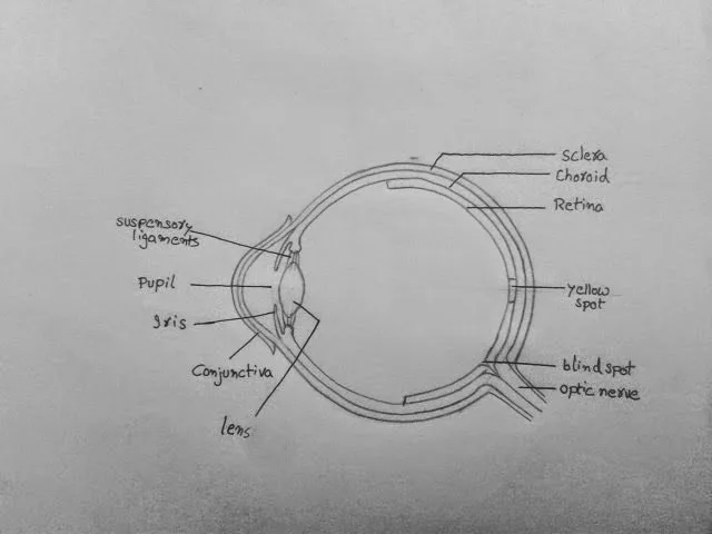
Sclera (Sclerotic layer): The eye’s strong outside coating is called the sclera. It creates the “White of the eye.” It safeguards and preserves the form of the eyeball and is made up of thick fibrous tissue.
Cornea: The cornea, a translucent window formed by the center covers one-sixth of the sclera, is colorless and transparent. The cornea permits light to flow through it and, by refraction, directs the light to the retina.
Conjunctiva: Thin, transparent, cell layer covering the cornea. A single layer of stratified squamous epithelium makes up this structure.
The middle vascular layer: It is made up of 3 parts.
Choroid: Located just below the sclera. It contains many bloods vessel and pigmented cells. The choroid’s dark tint is caused by a high concentration of melanin that is pigmented in its cells. It doesn’t reflect light back out of the eyeball because it absorbs it.
Ciliary body: Ciliary body contains smooth muscles called ciliary muscles in the eye. Ciliary muscles alter the shape of lens for near or far vision. This mechanism is called accommodation.
Iris: The iris has radial and circular muscles, which determine the shape of pupil. Pupil is the area through which light enter the eye ball.
Retina or Inner layer
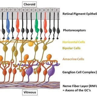
Retina is the inner layer which is highly photosensitive. It consists of two types of photoreceptor cells-rods and cones and photosensitive bipolar and ganglionic layers.
Rod cells: On the retina, there are 10–20 million rods. They create a black and white image when there is minimal light intensity dim light. They contain a pigment rhodopsin (visual purple) formed of Vitamin A. Rhodopsin is made up of 2 chemicals – opsin (a protein) and retinal (a derivative of Vitamin A).
Cone cells: On the retina, there are 7 million cones. They function in bright light and differentiate colors. They have iodopsin, a photosensitive pigment (Violet in color). Iodopsin comes in three distinct forms, each of which reacts to light of a different wavelength. There are red, green and blue sensitive cones.
Difference between Rod and Cone cells
| Parameter | Rod cells | Cone cells |
| Location | Located at the peripheral portion of retina | Located at the central portion of retina |
| Shape | Cylindrical and comparatively longer than cone cells | Comparatively shorter than rod cells |
| Narrower than cone cells | Wider than rod cells | |
| Number | More number of rod cells than cone cells | Number of cone cells are less than rod cells |
| Average number of rod cells in human is 120 million | Average number of cone cells in human is 6 million | |
| Sensitivity | Extremely sensitive to dim light of low intensity light | Sensitive to bright or high intensity light |
| Rod cells can be activated by even single photon of light | Large number of photons is required to trigger cone cells | |
| Vision | Rod cells help in scoptic vision (low light vision) and night vision | Cone cells help in photopic vision (high light vision) or day vision |
| Fovea | Absent in fovea | Concentrated in fovea |
| Photosensitive pigment | Photosensitive pigment is called rhodopsin or visual pigment | Photosensitive pigment is called Iodopsin or visual violet |
| Regenerative power | Rod cells possess very rapid regenerative power | The regenerative power of cone cells is slow. |
Rods and cones are both present in retina. The fovea centralis, also known as the yellow spot or macula lutea, is a tiny depression on the retinal wall that exclusively houses cones. This region is the most light-sensitive, and it creates an enlarged picture. It creates the most acute or crisp eyesight.
At a location known as the blind spot, the optic nerve penetrates the retina. There are no cones or rods in it. Since it is least sensitive to light, there is no formation of an image.
Lens
A transparent, elastic, biconvex and fibrous crystalline lens is present behind the iris. Ciliary muscles control the thickness of lens and its power of accommodation. The lens separates the eyeball into two chambers: a. Aqueous chamber and b. Vitreous chamber
Aqueous chamber: It is a little space between the cornea and the lens. It contains aqueous humor, a watery fluid. The lens, cornea, and supporting lens are all nourished by it. Additionally, it refracts light to focus on the retina.
Vitreous chamber: It is a large chamber between the lens and retina. It is filled with gelatinous fluid called vitreous humor. It helps to support retina. It also refracts the light rays to focus on retina.
Working Mechanism of Eye:
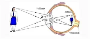
The conjunctiva, cornea, aqueous humor, lens, and vitreous humor are all tissues that the object’s light rays pass through in that order. All these structures refract the light such that it falls on the retina. This is called focusing. Maximum focusing is done by the cornea and the lens. The light then falls on the retina.
The light is received by the photoreceptors – rods and cones, on the retina. The absorbed light activates the pigments present in the rods and cones. The pigments are present on the membranes of the vesicles. Thus, the light is then converted into action potential in the membrane of vesicles. These travel as nerve impulses through the rod and the cone cells and reach the synaptic knob.
From here, impulses travel to the ganglions, then to the bipolar nerve cells, and finally to the optic nerves. Thus, about one million neurons of the optic nerve transmit the nerve impulses produced in the retina to the brain.
The occipital lobe in the rear of the brain regulates vision. After the information is processed, the picture is seen. The retina forms an inverted picture. The brain, however, causes us to view the image upright. Despite the fact that vision depends on the eyes, any injury to the optic nerves also impairs vision.