The human ear is a sensory organ designed specifically for sound detection and balance maintenance. Structure and Working Mechanism of Human Ear will be described in this article.The auditory nerve connects it to the brain. There are three sections to it.
- External Ear
- Middle Ear
- Internal Ear (the membranous labyrinth)
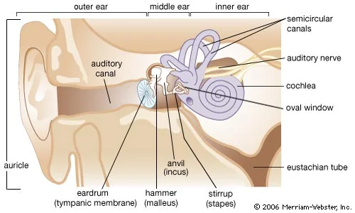
External Ear
The external ear consists of two different parts: a. Pinna (ear lobe) b. Auditory canal (meatus).
Pinna: It is the outer ear’s fleshy portion. It is supported by a flexible cartilage. It has a cup-like form and expands to capture sound waves.
Auditory canal (meatus): It’s a little tube called the auditory canal. Its length is roughly 2.5 cm. Wax glands that produce ear wax are located in its inner portion. The fragile eardrum is shielded by hair and wax from dust and microscopic insects.
The eardrum, also known as the tympanic membrane, is located at the end of the auditory canal. It serves as a boundary between auditory canal and middle ear.
Middle Ear
A little chamber located inside the temporal bone is the middle ear. Tympanic cavity refers to the air-filled chamber. Three ear ossicles, also known as tympanic bones, make up the hollow.
Malleus: Hammer shaped. It remains attached to the ear-drum.
Incus: Anvil shaped. It lies between malleus and stapes.
Stapes: Stirrup shaped. It is attached to the fenestra ovalis. Smallest bone of human body.
Ligaments that support these bones allow them to move freely as they transport sound from the tympanic membrane to the inner ear. These ossicles communicate the ear drum with internal ear through fenestra ovalis.
The Eustachian tube connects the middle ear with pharynx (throat). This connection equalizes the pressure in the middle ear with that of the outer atmosphere. When eustachian tube becomes blocked by cold, the inner and outer pressure do not equalize. If the pressure becomes great enough, the ear drum may burst.
Internal Ear
The internal human ear is the complex and delicate part of the ear. It is also called membranous labyrinth. It is enclosed in a bony labyrinth (cavity) containing perilymph fluid.
It consists of two different parts: a. Cochlea b. Vestibule
Cochlea: Cochlea is the fluid filled spiral tube. It looks like a tiny snail-shell. Its inner surface is lined with nerve endings. These nerve endings are highly sensitive to the vibration of fluid. All these nerve endings join the auditory nerve. This nerve leads from the cochlea to the brain.
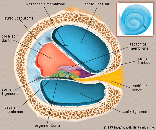
The tube of cochlea is divided longitudinally into three canals by two delicate membranes called Reisner’s membrane and basilar membrane. The three canals are:
Scala vestibuli: It is connected with tympanic cavity through fenestra ovalis filled with perilymph fluid.
Scala media: The scala media is the middle cells canal. It is filled with endolymph. The floor of the scala media is called the basilar membrane and its roof is called the Reisner’s membrane. The basilar membrane is provided with a sensory spot responsible for hearing, called organ of corti. The later is provided with the receptor cells, called sensory hair cells. The hair cells are provided with numerous hairs like structures at the free end. The scala vestibuli and scala tympani are filled with perilymph, communicate through helicotrema.
Scala tympani: It is connected with tympanic cavity through fenestra rotunda filled with perilymph.
Functions:
- Organ of corti conducts the sound waves thus associated with hearing.
- Vestibuli keeping a sense of balance and equilibrium.
Working Mechanism of Human Ear (Hearing)
Hearing is one of the main functions of ear that detects sound waves or vibrations in the air. Besides detecting, it also judges the direction and loudness of sound.
The pinna (auricle) is specially built to concentrate the sound waves towards the auditory meatus. The sound waves vibrate the tympanic membrane. The vibration from the tympanic membrane is then transmitted through the middle ear (tympanic cavity) by ossicles to reach the membrane of fenestra ovalis.
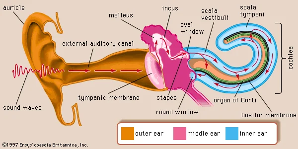
The human ear works in this direction:
Sound waves- Pinna and auditory meatus- tympanic membrane- ear ossicles- fenestra ovalis- perilymph of membranous labyrinth- cochlear duct- Scala media (endolymph)- Reissner’s membrane- basilar membrane- Organ of corti- Auditory nerves- Brain- Hearing
Equilibrium or Balance
The utriculus, sacculus and semicircular canals of the vestibule are the structure of our sense of balance or equilibrium. It is if two types:
Dynamic equilibrium: It is concerned with the balance of the body during locomotion and movement. Cristae found in the ampullae of semicircular canals are responsible for detection of rotational movements of the body. As there are three semi circular canals, three cristae detect the difference in position and direction. The endolymph of semicircular canals moves with the movement of the body. It stimulates the sensory hair cells of cristae to produce nerve impulses which are sent to the brain via auditory nerve.
Static equilibrium: It is concerned with the static equilibrium and linear acceleration of the body. Maculae found in the vestibules of utriculus and saccules are responsible for static equilibrium. Regions of the walls of the utricle and saccule, called maculae contains receptor cells. These cells have hair like processes embedded in a gelatinous mass that contains granules of Calcium carbonate. This mass is called Otoconium. Otoconia respond to the pull of gravity acting in right angles to the earth surface and are mainly responsible for detecting the direction of movement of head with respect to gravity.
Major Ear problems
Ear infection (otitis media)
Ear infections most commonly occur in your middle ear. Otitis media develops when bacteria and viruses become trapped in your middle ear. This type of infection is more likely to affect children than adults. Ear infection treatment usually involves antibiotics. In severe cases, ear tubes may be necessary.
Eustachian Tube dysfunction
The throat and middle ears are connected by the eustachian tubes. The eustachian tubes open when we yawn, sneeze, or swallow to balance the pressure inside of your ears. Eustachian tube dysfunction is the term used to describe when these tubes plug up. Tinnitus, distorted hearing, a feeling of fullness, and potential ear discomfort are among the symptoms.
Swimmer’s ear (otitis externa)
An ear canal infection known as swimmer’s ear is brought on by bacteria or fungus. This illness can be brought on by getting water in your ears.
Ruptured ear drum:
A ruptured eardrum occurs when the tympanic membrane becomes damaged. (Eardrum separates outer ear from middle ear.) A burst eardrum may result from an infection, trauma, loud noises, or foreign objects in the ears.
Learn more: