Cell division represents the fundamental biological process by which a parent cell divides to give rise to two or more daughter cells. This elegant mechanism serves as the primary means of reproduction for single-celled organisms, such as yeast and bacteria. Furthermore, in multicellular organisms—including humans—cell division acts as the engine behind growth, development, and tissue repair. It also facilitates the generation of specialized reproductive cells, specifically sperm and eggs.
Because life depends on the precise duplication of genetic material, the body maintains cell division as a tightly regulated process. When this regulation fails, the results can be catastrophic. Specifically, aberrant cell division often triggers pathological conditions, most notably cancer. This comprehensive guide explores the rich history of the field, the core mechanisms of the cell cycle, and the cutting-edge tools scientists utilize to study these microscopic phenomena.
A Brief History of Landmark Discoveries
The study of cell division has evolved significantly, moving from simple observations of “thread-like” structures to the complex molecular mapping of genetic switches.
The Birth of Cell Theory
The scientific journey began in the 1600s when Anton van Leeuwenhoek and Robert Hooke utilized early microscopes to reveal the invisible world of cells. However, it wasn’t until the 1830s that botanists Barthélemy Dumortier and Hugo von Mohl observed a critical phenomenon: new plant cells were created through the division of existing ones.
By 1838, Matthias Jakob Schleiden and Theodor Schwann synthesized these observations into the first two tenets of Cell Theory:
All living organisms are composed of one or more cells.
Cells function as the basic building blocks of all life.
Nearly twenty years later, a physician named Rudolf Virchow added the definitive third tenet, which stated that all cells arise from preexisting cells.
Defining Mitosis and Meiosis
In 1876, Walther Flemming observed cells dividing and noticed the separation of unique, thread-like structures. Consequently, he coined the term mitosis, derived from the Greek mitos (thread). Shortly thereafter, Edouard Van Beneden and Theodor Heinrich Boveri identified these threads as chromosomes. They also discovered centrosomes, the structures that organize the microtubules responsible for pulling chromosomes apart.
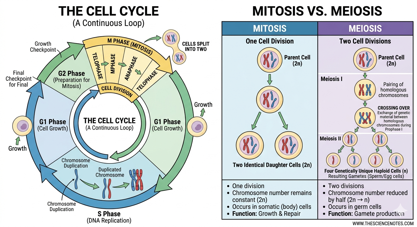
While mitosis produces identical daughter cells, researchers like Oscar Hertwig and August Weismann focused on meiosis. This specialized division involves one round of DNA replication followed by two rounds of division. As a result, meiosis produces gametes with half the number of chromosomes found in the parent cell.
The Molecular Revolution
During the latter half of the 20th century, the focus shifted toward regulation. In 1972, Leland Hartwell identified “cell division cycle” (cdc) genes in yeast. Following this, in 1983, Tim Hunt discovered cyclins, which are proteins that oscillate in abundance to trigger different phases of the cell cycle. Sir Paul Nurse later proved that these regulatory genes were highly conserved across species, including humans. Their collective work earned the Nobel Prize in 2001, highlighting the global importance of these biochemical switches.
Fundamental Questions in Modern Cell Biology
Today, researchers move beyond merely watching what happens; instead, they investigate how it is controlled. Several key questions currently drive the field:
1. What Regulates the Biochemical Switches?
Intracellular signaling pathways and genes govern the cell cycle through a series of checkpoints. Scientists are currently working to map the intricate molecules that act as “go” or “no-go” signals for DNA replication. Understanding these switches is essential for learning how cells maintain genomic stability.
2. How Do Extracellular Factors Influence Division?
Cells rarely exist in isolation. Instead, they respond to external chemical cues called mitogens. Biologists are striving to understand which specific external factors stimulate or inhibit division, as this knowledge could unlock new ways to control tissue regeneration.
3. What Drives Pathological Proliferation?
Abnormal cell division remains the primary hallmark of cancer. While we know that mutations in oncogenes initiate disease, many factors in tumor progression remain a mystery. Therefore, researchers are working tirelessly to reveal unknown proteins that cause “mitotic failure” and uncontrolled growth.
Essential Tools in Cell Division Research
To answer these complex questions, biologists employ a suite of sophisticated laboratory techniques designed to track cellular behavior.
Cell Cycle Analysis via Flow Cytometry
By using fluorescent dyes, scientists can determine which phase of the cycle a cell is in with high precision.
BrdU (Bromodeoxyuridine): This thymidine analog incorporates into DNA during the S phase (synthesis). Consequently, it labels only the cells actively replicating DNA.
Propidium Iodide (PI): This compound stains all DNA within a sample. Because cells in the G_2 phase have twice as much DNA as those in G_1, the intensity of the PI signal reveals the exact stage of each cell.
Live Cell Imaging and Time-Lapse Microscopy
Advances in imaging now facilitate the direct observation of division. Scientists can “tag” specific parts of a cell—such as the spindle fibers—using fluorescent proteins. Afterward, they use time-lapse microscopy to create a real-time record of the cellular machinery in action.
Quantifiable Tracking Dyes
To track how many times a cell has divided over several days, researchers use membrane-binding dyes. Each time a cell divides, the daughter cells receive exactly half of the parent’s dye. As the signal becomes dimmer, the diminishing fluorescence intensity allows scientists to identify different generations of cells within a mixed population.
Real-World Applications of Cell Division Studies
These techniques are not merely academic; rather, they are vital for medical and genetic breakthroughs.
Genetic Mutation Analysis
In organisms like Drosophila (fruit flies), scientists study how specific genetic mutations affect tissue development. By performing genetic crosses and analyzing the wing tissue, researchers can identify which genes are essential for healthy growth.
Drug Development for Cancer
Pharmaceutical researchers use fluorescence microscopy to test new chemotherapy candidates. For instance, in a recent experiment, scientists treated cancer cells with a drug called JP-34. The results showed that the drug forced the cells into mitotic failure and eventual death, proving its potential as a therapeutic agent.
Immunology and Proliferation Rates
Not all cells divide at the same speed. By using tracking dyes, immunologists have discovered that different types of immune cells proliferate at vastly different rates during an infection. This insight helps in the design of vaccines and treatments for autoimmune disorders.
Conclusion
From the early sketches of Robert Hooke to the high-speed laser scanning of modern flow cytometry, our understanding of cell division has expanded exponentially. It remains a process of incredible precision, serving as the bridge between generations of life. As we continue to uncover the molecular secrets of the cell cycle, we move closer to mastering the “switches” of life, offering hope for new treatments for cancer and a deeper understanding of our own biological origins.
Summary Table: Key Phases of the Cell Cycle
| Phase | Name | Primary Function |
| G_1 | Gap 1 | The cell grows and prepares for DNA synthesis. |
| S | Synthesis | The cell replicates its DNA to double the genetic material. |
| G_2 | Gap 2 | The cell continues to grow and checks DNA for errors. |
| M | Mitosis | The nucleus and cytoplasm physically divide into two cells. |