The human brain is a masterpiece of biological engineering, but its most critical features aren’t just the solid tissues. Deep within the folds of the cerebral cortex lies a hidden, fluid-filled network known as the ventricular system. These hollow cavities serve as the primary production site for cerebrospinal fluid (CSF), a clear, colorless liquid that acts as the brain’s internal ocean.
Without this complex plumbing system, the brain would collapse under its own weight or succumb to metabolic toxicity. In this comprehensive guide, we will explore the anatomy of the four ventricles, the physiology of the choroid plexus, and the medical conditions that arise when this delicate balance is disrupted.
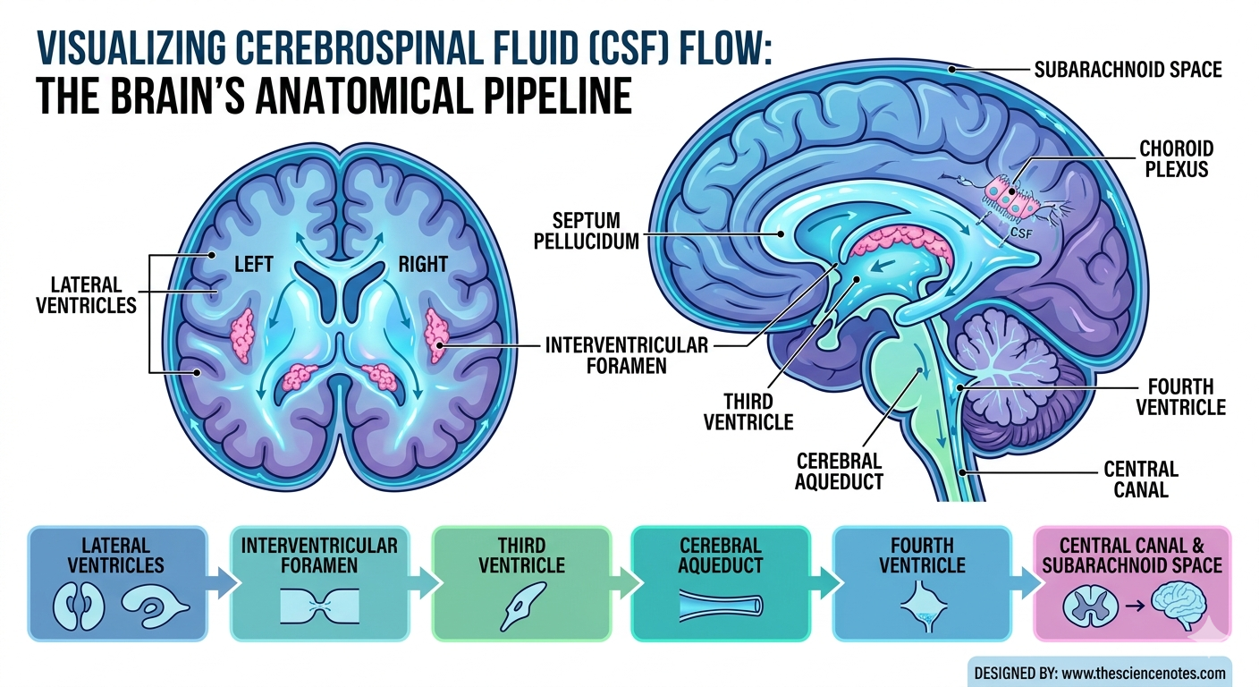
1. The Lateral Ventricles: The Largest Cavities
The two lateral ventricles represent the largest parts of the ventricular system. Each cerebral hemisphere houses one of these expansive, C-shaped chambers. Because they follow the developmental growth of the cerebrum, they reach into nearly every major lobe of the brain.
The Three Horns and Body
Anatomists divide the lateral ventricles into several distinct regions. When viewed from the side, each ventricle shows three “horns” that act as projections:
Anterior (Frontal) Horn: This section extends forward into the frontal lobe. It plays a role in surrounding the head of the caudate nucleus.
Body (Central Part): This is the main portion of the ventricle, located within the parietal lobe.
Inferior (Temporal) Horn: This section curves downward and forward into the temporal lobe, sitting near the hippocampus.
Posterior (Occipital) Horn: This projection reaches backward into the occipital lobe.
The Septum Pellucidum
The two lateral ventricles sit remarkably close together at the midline. However, they do not merge. Instead, a thin, triangular membrane called the septum pellucidum separates them. This vertical sheet of tissue acts as a clear partition between the left and right sides. If this membrane is absent, it often indicates a developmental neurological condition.
2. The Diencephalon’s Gateway: The Third Ventricle
As fluid moves out of the lateral ventricles, it enters the third ventricle. This is a narrow, slit-like cavity located in the center of the diencephalon.
The Interventricular Foramen
The communication between the lateral ventricles and the third ventricle happens through a tiny opening. Medical professionals call this the interventricular foramen (or the Foramen of Monro). Because these openings are so small, they are often the first sites to become blocked if a tumor or inflammation occurs.
Surrounding Structures
The third ventricle acts as a landmark for several vital brain structures. The left and right halves of the thalamus form its lateral walls. Meanwhile, the hypothalamus forms its floor and lower walls. This central location makes the third ventricle a critical point for neurosurgical procedures involving the deep brain.
3. The Lower Pathway: The Cerebral Aqueduct and Fourth Ventricle
To reach the lower parts of the central nervous system, CSF must pass through the brainstem. This pathway is the narrowest part of the entire system.
The Cerebral Aqueduct
The cerebral aqueduct (also known as the Aqueduct of Sylvius) is a long, canal-like structure. It runs through the midbrain, connecting the third and fourth ventricles. Because it is so slender—measuring only a few millimeters in diameter—it is highly susceptible to narrowing (stenosis).
The Fourth Ventricle
The fourth ventricle is a diamond-shaped cavity. It sits posterior to the pons and medulla oblongata and anterior to the cerebellum. This chamber serves as the final “staging area” for CSF. From here, the fluid exits through three small openings (the foramina of Luschka and Magendie) to surround the outer surface of the brain and spinal cord.
4. The CSF Factory: The Choroid Plexus and Ependymal Cells
The ventricles are not merely passive storage tanks; they are active chemical factories. They produce roughly 500 milliliters of cerebrospinal fluid (CSF) every single day. Since the total volume in the system is only about 150 milliliters, the body must constantly recycle and replace this fluid.
The Blood-CSF Barrier
A specialized structure called the choroid plexus lines the walls of all four ventricles. This network consists of folded capillaries surrounded by ependymal cells. These cells are joined by “tight junctions,” creating a highly selective Blood-CSF Barrier.
This barrier performs three essential tasks:
Filtration: It allows water and small molecules to pass from the blood into the brain.
Exclusion: It keeps harmful bacteria, large proteins, and most toxins out of the brain’s internal environment.
Secretion: Ependymal cells actively transport ions like sodium and chloride into the ventricles to “pull” water in, creating the fluid.
5. The Vital Functions of Cerebrospinal Fluid
The fluid produced within these ventricles is essential for survival. It performs several mechanical and physiological roles that keep the central nervous system (CNS) healthy.
Buoyancy and Protection
The human brain is heavy and soft. Without support, its own weight would tear the delicate nerves and blood vessels at its base. CSF provides buoyancy, reducing the brain’s effective weight from 1,400 grams to just 50 grams. This “floating” effect allows the brain to survive sudden impacts and mechanical shocks.
Chemical Homeostasis
The brain requires a very specific chemical environment to function. CSF regulates the concentration of electrolytes like potassium and calcium. This balance is necessary for neurons to fire electrical signals correctly. If the chemistry shifts even slightly, it can lead to seizures or confusion.
The Glymphatic System and Waste Removal
Recent research highlights the “glymphatic system.” During sleep, the flow of CSF between brain cells increases significantly. This process flushes out metabolic waste, including beta-amyloid, a protein associated with Alzheimer’s disease. Therefore, healthy ventricular flow is a key component of long-term cognitive health.
6. Clinical Significance: When the System Fails
Understanding the ventricles is vital for diagnosing many neurological disorders. When the production, flow, or reabsorption of CSF is disrupted, the results can be life-threatening.
Hydrocephalus: “Water on the Brain”
Hydrocephalus occurs when CSF builds up within the ventricles. This usually happens for one of three reasons:
Obstructive Hydrocephalus: A physical blockage (like a tumor or narrow aqueduct) stops the flow.
Communicating Hydrocephalus: The fluid flows correctly but is not reabsorbed into the bloodstream.
Overproduction: In rare cases, the choroid plexus produces too much fluid.
In infants, this causes the head to enlarge. In adults, it increases intracranial pressure, leading to headaches, vision loss, and cognitive decline.
Ventriculitis and Meningitis
Inflammation of the ventricular lining is known as ventriculitis. This often occurs alongside meningitis. Because the ventricles connect directly to the spinal cord, infections in this fluid can spread rapidly throughout the entire nervous system.
Age-Related Ventricular Enlargement
As we age, brain tissue naturally shrinks (atrophy). To fill the resulting empty space, the ventricles enlarge. Doctors use the size of the lateral ventricles on MRI scans to track the progression of neurodegenerative diseases like dementia.
The Anatomical Pipeline: A Flow Summary
To help students visualize the circulation, we can summarize the path as follows:
Lateral Ventricles: Production begins via the choroid plexus.
Interventricular Foramen: Fluid enters the midline.
Third Ventricle: More fluid is added in the diencephalon.
Cerebral Aqueduct: Fluid travels through the narrow midbrain path.
Fourth Ventricle: Fluid prepares to exit the internal brain.
Subarachnoid Space: CSF flows around the exterior of the brain and spinal cord.
Arachnoid Granulations: Fluid is reabsorbed back into the venous blood.
Conclusion
The ventricular system is far more than just “holes in the brain.” It is a dynamic, highly regulated environment that provides the mechanical and chemical support necessary for human thought and movement. By acting as a cushion, a nutrient delivery system, and a waste removal service, the ventricles ensure that our most complex organ remains in peak condition. Whether you are a student of biology or simply curious about human health, understanding these fluid-filled spaces is essential to appreciating the miracle of the human mind.