The cerebrum is the crown jewel of the human central nervous system. As the largest and most highly developed part of the brain, it occupies the majority of the cranial cavity. It serves as the command center for everything from basic sensory perception to complex philosophical thought. While we often think of the brain as a single unit, the cerebrum is actually a deeply partitioned structure. Its unique design—consisting of folds, grooves, and distinct lobes—allows it to process a massive amount of information with incredible efficiency.
In this comprehensive guide, we will explore the anatomical landscape of the cerebrum. We will examine the functional roles of the five lobes, the importance of the gyri and sulci, and how the brain integrates with the protective structures of the skull.
1. The Architecture of the Cerebral Cortex
The cerebrum consists of two primary regions: the outer cerebral cortex and the underlying white matter. The cortex is a thin layer of “grey matter,” usually only 2 to 4 millimeters thick. However, this thin layer contains billions of neurons that form the basis of our consciousness.
Understanding Gyri and Sulci
The most striking feature of the cerebrum is its wrinkled appearance. This is not a random developmental byproduct. Instead, it is a brilliant evolutionary adaptation to increase surface area within the limited space of the skull.
Gyri (singular: Gyrus): These are the elevated ridges or “bumps” on the brain’s surface.
Sulci (singular: Sulcus): These are the shallow grooves or “valleys” that separate the gyri.
By folding the cortex into these gyri and sulci, the human brain triples its surface area. Consequently, this allows for a higher density of neurons, which directly correlates with our advanced cognitive abilities compared to other mammals.
2. The Hemispheres and the Great Fissure
A deep, prominent groove called the medial longitudinal fissure divides the cerebrum into the left and right cerebral hemispheres. While these two halves look identical, they are functionally distinct—a concept known as hemispheric lateralization.
The left hemisphere generally dominates language, logical reasoning, and mathematical calculations. Conversely, the right hemisphere often excels in spatial visualization, facial recognition, and artistic awareness. Despite these differences, the two halves communicate constantly through a thick bundle of white matter fibers called the corpus callosum.
3. Mapping the Five Lobes of the Cerebrum
Specific large and distinct sulci act as anatomical boundaries, dividing each hemisphere into five different lobes. Four of these lobes are visible on the exterior, while the fifth is hidden deep within the brain’s folds.
The Frontal Lobe: The Executive Suite
Located at the very front of the cerebrum, the frontal lobe is the center for “executive functions.” This includes planning, decision-making, and personality expression. Furthermore, it contains the precentral gyrus, which acts as the primary motor cortex. This area sends the final electrical signals that tell your muscles to move.
The Parietal Lobe: The Sensory Processor
Positioned behind the frontal lobe, the parietal lobe handles the processing of sensory information. It interprets signals related to touch, pressure, temperature, and pain. It houses the postcentral gyrus, known as the primary somatosensory cortex. This region maps every inch of your body to a specific part of the brain.
The Temporal Lobe: The Auditory Center
Situated on the sides of the brain, near the ears, the temporal lobe is primarily responsible for hearing. It also plays a massive role in memory formation and language recognition. The lateral sulcus separates this lobe from the frontal and parietal regions above it.
The Occipital Lobe: The Visual Hub
Located at the posterior (back) of the cerebrum, the occipital lobe is dedicated almost entirely to vision. It receives raw data from the eyes and translates it into the shapes, colors, and motions we perceive as the world around us.
The Hidden Gem: The Insula
The insula is the smallest and most elusive lobe. It is present deep within the lateral sulcus. To see it, an anatomist must physically pull the temporal and frontal lobes apart. The insula is involved in diverse functions, such as the perception of pain, the sense of taste (gustatory perception), and emotional awareness.
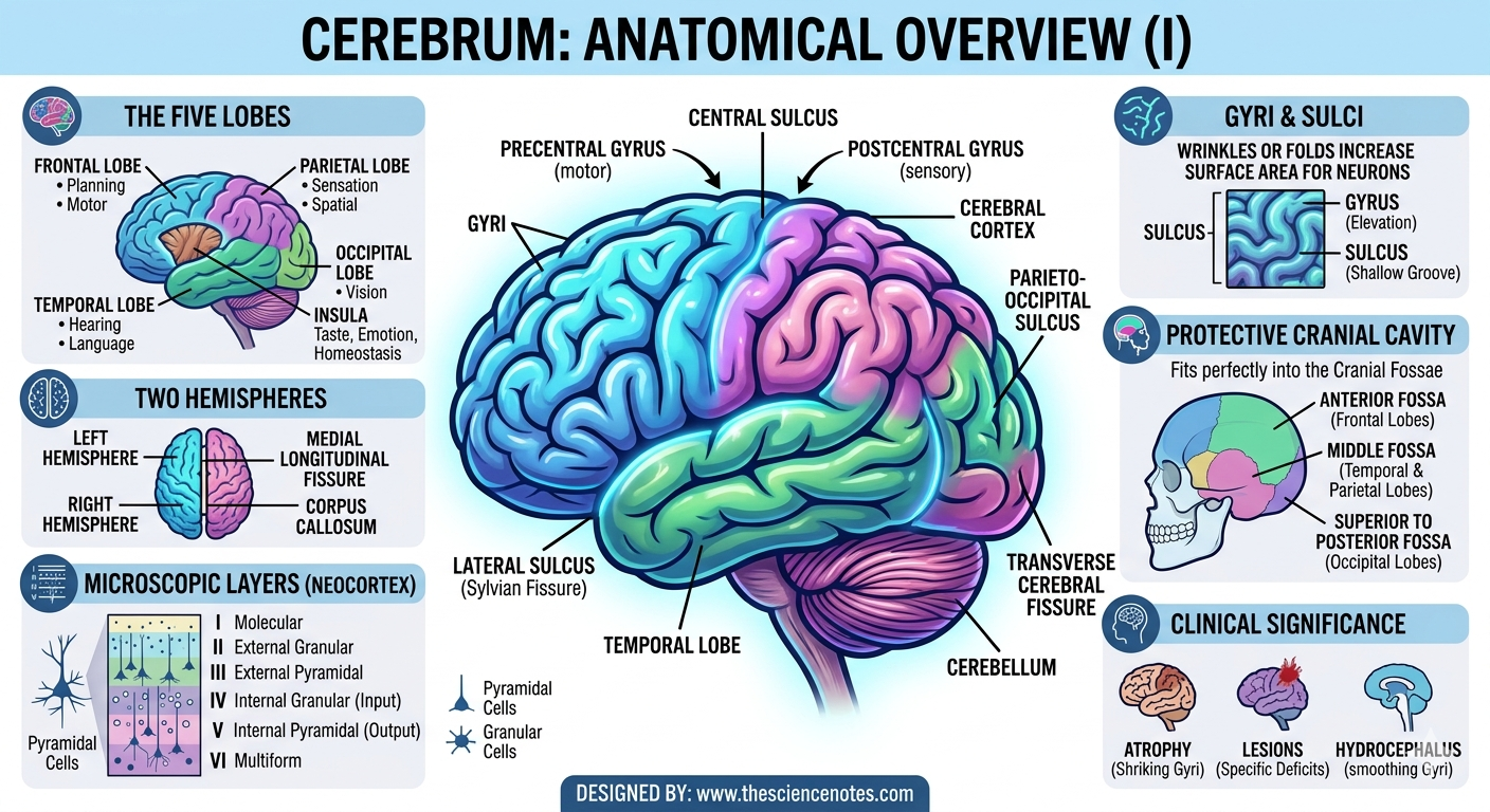
4. Major Landmarks: The Sulci that Define the Brain
To navigate the cerebrum, one must understand the major “roads” or landmarks that separate these functional zones.
The Central Sulcus: This is perhaps the most important groove in the brain. It separates the frontal lobe from the parietal lobe. Most importantly, it creates the physical divide between movement (motor control) and feeling (sensory perception).
The Lateral Sulcus (Sylvian Fissure): This deep groove acts as a boundary between the temporal lobe below and the frontal and parietal lobes above.
The Parieto-Occipital Sulcus: This groove marks the transition between the parietal and occipital lobes. It is clearly visible on the medial (inner) surface of the brain.
The Transverse Cerebral Fissure: This deep separation marks the end of the occipital lobe. It is crucial because it separates the cerebrum from the cerebellum (the “little brain”) situated beneath it.
5. Microscopic View: The Layers of the Cortex
While the macroscopic view shows lobes and gyri, the microscopic view reveals a sophisticated cellular organization. The cerebral cortex is typically organized into six horizontal layers (neocortex).
Molecular Layer: Mostly contains nerve fibers and few cells.
External Granular Layer: Involved in communication between different cortical areas.
External Pyramidal Layer: Contains cells that send signals to other parts of the cortex.
Internal Granular Layer: The primary “input” layer, receiving sensory data from the thalamus.
Internal Pyramidal Layer: The “output” layer, sending motor signals down to the spinal cord.
Multiform Layer: Connects the cortex back to the thalamus.
This layered structure ensures that the brain can receive, process, and send information in an orderly, highly efficient fashion.
6. Protection: How the Skull Cradles the Cerebrum
The cerebrum is a soft, fragile organ that requires substantial protection. The skull’s internal floor is not flat; it is shaped into three “basins” called cranial fossae. Each lobe of the brain fits perfectly into these depressions:
Anterior Cranial Fossa: Cradles the frontal lobes.
Middle Cranial Fossa: Houses the temporal and parietal lobes.
Posterior Cranial Fossa: The occipital lobes sit superior to this area, which primarily houses the cerebellum and brainstem.
This tight fit minimizes the “sloshing” of the brain during movement. Furthermore, the meninges (protective membranes) and cerebrospinal fluid provide additional layers of security against mechanical trauma.
7. Clinical Significance: When Anatomy Changes
Understanding the standard anatomy of the cerebrum allows doctors to identify neurological issues.
Atrophy: In diseases like Alzheimer’s, the gyri become thinner and the sulci become wider and deeper as brain cells die.
Lesions: Damage to a specific gyrus can result in very specific deficits. For example, damage to the precentral gyrus can cause paralysis on the opposite side of the body.
Hydrocephalus: If fluid builds up in the brain, it can press the cerebrum against the skull, smoothing out the gyri and damaging the cortex.
Conclusion
The cerebrum is a masterpiece of biological architecture. From the complex folding of the gyri and sulci to the strategic placement of the five lobes, every detail serves a functional purpose. Its design maximizes processing power while ensuring the organ remains protected within the cranial cavity. By understanding this anatomical overview, we gain a deeper appreciation for the miracle of human consciousness and the physical structures that make our thoughts and actions possible.