While the primary motor and sensory areas of the brain handle the “raw data” of our existence—executing a movement or registering a sound—the true magic of the human mind happens in the association areas. These regions of the cerebral cortex do not have a single, dedicated sensory or motor function. Instead, they act as the brain’s great integrators.
By weaving together information from various lobes, memories of the past, and emotions of the present, association areas enable higher cognitive processes. Without them, we could see an object but not know what it is; we could hear a voice but not understand the words. In this guide, we will explore the complex landscape of these areas, from the centers of language to the high-level decision-making hubs of the frontal lobe.
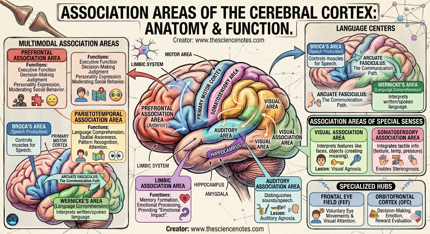
1. The Master Planners: Multimodal Association Areas
Multimodal association areas receive input from multiple sensory modalities and create a complete “picture” of our environment. They allow us to give meaning to the world around us.
The Prefrontal Association Area (Anterior Association Area)
Located in the anterior portion of the frontal lobe, this is arguably the most evolved part of the human brain. It is the seat of executive function.
Role: It manages complex learning abilities, recall, judgment, and abstract thinking.
Connections: It maintains numerous connections with the thalamus, hypothalamus, and the limbic system.
Function: Because it connects with emotional centers, it helps moderate social behavior. It allows us to weigh the consequences of our actions, suppress impulses, and plan for the future.
The Parietotemporal Association Area (Posterior Association Area)
Found at the junction of the parietal and temporal lobes, this area is essential for providing us with a “sense of place.”
Role: It integrates input from the primary auditory, visual, and somatosensory cortices.
Function: It is crucial for spatial awareness and recognizing patterns. It allows you to understand that the various sensory inputs you are receiving—the smell of coffee, the sight of a mug, and the heat on your hand—all belong to a single coherent experience.
The Limbic Association Area
Situated in the medial part of the temporal lobe, this area bridges the gap between thought and feeling.
Role: It connects directly with the hippocampus and amygdala.
Function: It is essential for memory formation and emotional processing. It provides the “emotional impact” that makes a scene important to us, helping us decide what is worth remembering.
2. The Language Centers: Broca’s and Wernicke’s Areas
Language is one of the most complex tasks the brain performs, requiring a precise handoff between production and comprehension. These two areas are usually located in the left hemisphere (the dominant hemisphere for language in most people).
Broca’s Area: The Architect of Speech
Located in the posterior part of the left inferior frontal gyrus, Broca’s area is the “motor” for language.
Function: It plans the sequence of muscle contractions needed to form words. It connects to the primary motor cortex to execute these plans.
Clinical Significance: Damage results in Broca’s Aphasia. Patients know what they want to say but struggle to physically produce the words. Speech is slow and labored, though they can still understand others.
Wernicke’s Area: The Interpreter of Meaning
Situated in the superior temporal gyrus, Wernicke’s area is the “translator.”
Function: It interprets spoken and written language. It allows us to recognize that a specific sound corresponds to a specific concept.
The Connection: The arcuate fasciculus, a bundle of nerve fibers, connects Broca’s and Wernicke’s areas. This allows us to hear a word, understand it, and formulate a spoken response.
Clinical Significance: Damage results in Wernicke’s Aphasia. Patients speak fluently, but their words are nonsensical (“word salad”). Furthermore, they usually cannot understand what is being said to them.
3. Association Areas of the Special Senses
While the primary cortices detect basic features (like a line or a pitch), the sensory association areas interpret those features into recognizable objects.
Visual Association Area
Located in the occipital lobe, surrounding the primary visual cortex (V1).
Process: V1 sees edges and colors; the association area sees a face or a car.
Visual Agnosia: If this area is damaged, a person might see a set of keys but be unable to identify them as “keys” until they pick them up and hear them jingle.
Auditory Association Area
Found in the temporal lobe, this area interprets the “meaning” of sounds.
Process: It allows you to distinguish between a doorbell, a dog barking, and a human voice.
Auditory Agnosia: Damage means a person can hear sounds but cannot interpret them. A symphony might sound like random noise.
Somatosensory Association Area
Situated in the parietal lobe, posterior to the postcentral gyrus.
Process: It integrates tactile information like texture, temperature, and pressure.
Stereognosis: This area allows you to reach into your pocket and identify a coin simply by feeling its shape and weight without looking at it.
4. Specialized Recognition and Control Hubs
Beyond the broad lobes, specific “patches” of the cortex are dedicated to highly specialized tasks.
The Facial Recognition Area (Fusiform Face Area – FFA)
Located in the fusiform gyrus of the temporal lobe, this region is a specialist.
Role: It distinguishes faces from other objects.
Mechanism: It works with the Occipital Face Area (OFA) to identify who a person is, while other areas process their expression or gaze.
Prosopagnosia: Damage to the FFA leads to “face blindness,” where individuals cannot recognize even their own family members by sight.
The Orbitofrontal Cortex (OFC)
Sitting just above the eye sockets in the frontal lobe, the OFC is the center for reward and value.
Role: It helps us make decisions based on stimuli. For example, it evaluates the “reward” of eating a piece of chocolate.
Function: It helps us respond to the emotional content of visual cues, such as a partner’s facial expression, and adjust our behavior accordingly.
The Frontal Eye Field (FEF)
This region in the frontal cortex manages our “visual search.”
Role: It controls voluntary, rapid eye movements (saccades).
Function: It allows us to prioritize relevant information. When you look for a specific name on a long list, the FEF is directing your gaze and managing your attention.
5. Integration and Higher Cognitive Processes
The true power of the association areas lies in their connectivity. They do not work in isolation. The brain uses association fibers to communicate within the same hemisphere and commissural fibers (like the corpus callosum) to share data between the left and right sides.
Learning and Memory
When you learn a new skill, the prefrontal cortex coordinates the focus, while the sensory association areas store the “patterns” of that skill. Over time, these pathways become more efficient, moving from conscious effort to automatic “muscle memory.”
Decision-Making and Emotion
The link between the prefrontal association area and the limbic system is what makes us human. It allows us to temper our emotions with logic. Consequently, damage to these connections often results in radical personality changes, as seen in the famous case of Phineas Gage.
Conclusion
The association areas of the cerebral cortex represent the pinnacle of neurological complexity. By acting as the bridge between “feeling” and “doing,” they allow us to interpret the world, communicate through language, and make complex decisions. From the spatial awareness of the parietal lobe to the moral judgment of the prefrontal cortex, these regions are the physical structures that house our identity, memory, and intelligence.