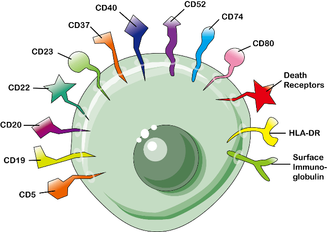
Clusters of Differentiation (CD) Antigens are the surface marker molecules expressed on the surface of cells of immune system (leukocytes) which show various activation or inactivation phases or several lineage-specific differentiation stages.
Immune cells produce complex variants of CD antigens on their cell surfaces during lymphocyte development; some of these antigens are lost, while others are gained at different phases through contacts with antigen presenting cells (APC) or with immune system cells.
They perform important roles in adaptive immunity, immunological cell-cell communication, and microenvironment sensing.
CD antigens are mostly membrane proteins that may be identified by where they are found within or on the surface of the phospholipid bilayer.
CD molecules frequently function as receptors or ligands—molecules that activate receptors—important to the cell. Typically, a signal cascade is started, changing the cell’s behavior. Certain CD proteins serve other purposes, such as cell adhesion, and are not involved in cell signaling. There are over 250 different types of proteins. Therefore, they do not belong to particular class of molecules.
Anti-leukocyte monoclonal antibodies are frequently used to identify leukocyte cell surface molecules (mAbs). The cell surface immunophenotypes of many leukocyte subpopulations, including the functionally separate mature lymphocyte subpopulations of B-cells, helper T-cells (TH), cytotoxic T-cells (TC), and natural killer (NK) cells, may be mapped using various mAb combinations.
CD Nomenclature
The Human Leukocyte Differentiation Antigens (HLDA) 1st International Workshop and Conference developed and defined the CD nomenclature. This approach was created to categorize the many monoclonal antibodies (mAbs) that were produced by several laboratories throughout the world against epitopes on the surface molecules of leukocytes (white blood cells).
This provided common universal language that allowed the scientific community to communicate.
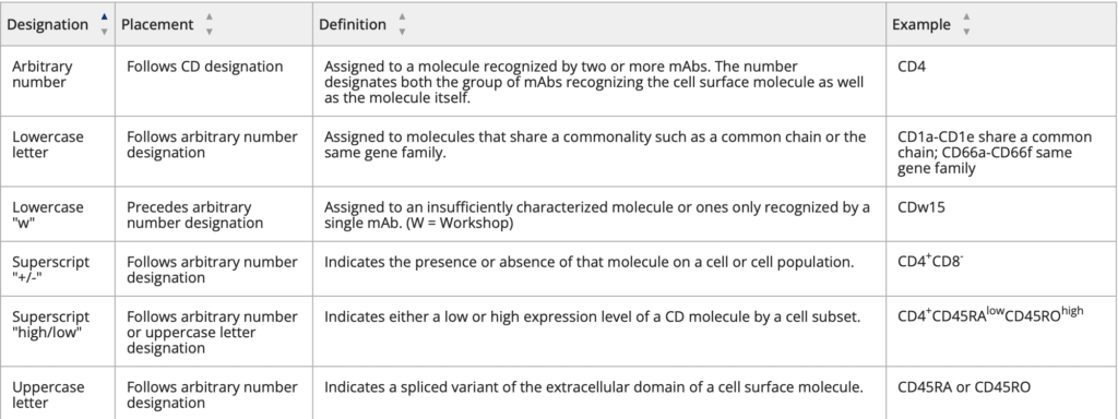
Cluster of Differentiation Rules
Source: https://www.aatbio.com/catalog/cd-cluster-of-differentiation-cell-surface-markers
How does the cluster of differentiation work?
The cluster of differentiation procedure, often known as CD, is used to find and analyze cell surface chemicals that serve as targets for cell immunophenotyping. In terms of physiology, CD antigens can function in a variety of ways, frequently serving as crucial receptors or ligands (a substance that activates a receptor). Typically, a signal cascade is started, changing the cell’s behavior. Certain CD antigens serve various purposes outside of contributing to cell signaling, including cell adhesion, cell activation, and cell inhibition.
In immunophenotyping, the CD antigens are frequently utilized as cell markers, allowing cells to be classified according to the molecules that are found on their surface. These markers are frequently employed to link cells to certain immunological activities. While it is rare to identify populations with only one CD molecule (although there are a few examples), combining markers has made it possible to define immune system cell types with extremely precise specifications.
Flow cytometry is one approach for cell sorting that makes use of CD antigens.
Immunophenotyping
Most often, CD antigens are found to identify immune system cell types and subpopulations. a technique that employs antibodies to recognize cells depending on the kinds of antigens or markers on their surface. This procedure is used to identify certain leukemia, lymphoma, and immune system cell types. Immunophenotyping frequently makes use of Clusters of Differentiation (CD) markers.
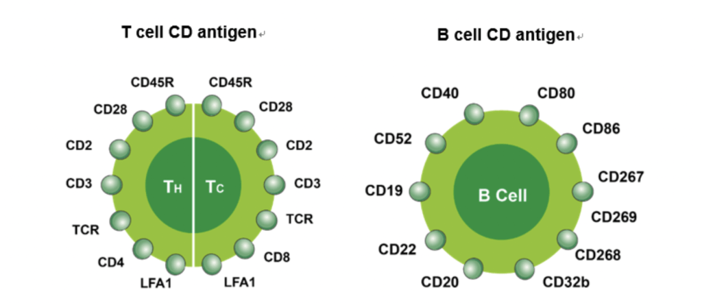
Since immune cells produce CD antigens at different phases of development or cell activation, they are useful indicators for distinguishing leukocyte populations and subpopulations, a process known as immunophenotyping. For instance, CD3 is a T cell co-receptor that is uniquely expressed on the surface of all mature T cells, whereas CD4 and CD8 antigens are expressed on the surfaces of T helper cells and cytotoxic T cells, respectively.
For instance, cells that express CD38 but not CD19 and CD20 are often indicative of multiple myeloma. Because of the variable levels and kinds of expression of CD antigens, it is possible to clearly identify a specific cell type or subtype using multiparameter flow cytometric analysis using two or more combinations of fluorescently labeled CD antibodies.
| Type of cell | CD markers |
| Stem cells | CD34+, CD31-, CD117 |
| all leukocyte groups | CD45+ |
| Granulocyte | CD45+, CD11b, CD15+, CD24+, CD114+, CD182+ |
| Monocyte | CD4, CD45+, CD14+, CD114+, CD11a, CD11b, CD91+, CD16+ |
| T lymphocyte | CD45+, CD3+ |
| T helper cell | CD45+, CD3+, CD4+ |
| T regulatory cell | CD4, CD25, FOXP3 (a transcription factor) |
| Cytotoxic T cell | CD45+, CD3+, CD8+ |
| B lymphocyte | CD45+, CD19+, CD20+, CD24+, CD38, CD22 |
| Thrombocyte | CD45+, CD61+ |
| Natural killer cell | CD16+, CD56+, CD3-, CD31, CD30, CD38 |
CD Markers in Solid Tumors
A very broad group of membrane proteins known as CD antigens are mostly expressed on the surface of leukocytes as well as other cell types like endothelial, stem, and dendritic cells. For cell sorting, phenotyping, and the identification of blood tumors, CD markers-specific antibodies are frequently utilized.
A common technique in cancer diagnosis, screening, and research is the use of antibodies to identify tumor antigens. Using tumor antigens-specific antibodies, tumors can be diagnosed and their prognosis assessed when there are high levels of tumor antigens in a patient’s blood, urine, or tissue. The prostate specific antigen used in the diagnosis of prostate tumor is the most extensively utilized tumor marker of this type.
Moreover, CD markers have taken on a key role in the therapy of tumors. Several therapeutic antibody medications are intended to target cells with a certain CD marker (e.g, rituximab to CD20 for lymphomas and leukemia treatment; alemtuzumab to CD52 for chronic lymphocytic leukemia and T-cell lymphoma treatment).
CD4 and CD8 Markers
Most thymocytes and almost two-thirds of peripheral blood T cells, which make up CD8- cells, express CD4. Monocytes and macrophages express CD4 in humans and rats but not in mice.
CD8 is expressed on most thymocytes and approximately one-third of peripheral blood T cells, which constitute the CD4- cells. CD8αβ heterodimers are expressed only on TCRαβ cells whereas CD8α homodimers can be expressed on αβ and γδ T cells and some NK cells.
In addition to MHC Class II antigens, CD4 is an auxiliary molecule that aids T lymphocytes in their detection of foreign antigens.
For the purpose of recognizing antigens, CD8 collaborates with MHC Class I-restricted TCRs.
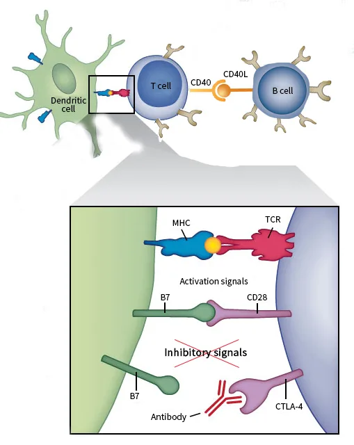
Learn More on T cell Signaling
Questions and Answers
When does T cells recognize antigen?
- When they are shown on antigen-presenting cells’ (APC) surfaces.
What is Antigen presenting cells (APC)?
- APCs are a kind of immune cells with the ability to process and display antigens so that T cells may recognize them and begin the adaptive cellular immunity responses.
References
- https://www.creativebiomart.net/Cluster-of-Differentiation.htm
- https://biosci.mcdb.ucsb.edu/immunology/Cells-Organs/CD-antigens.htm
- https://www.sinobiological.com/research/cd-antigens/clusters-of-differentiation
- https://wiki.clinicalflow.com/cluster-differentiation-cd-markers
- https://www.aatbio.com/catalog/cd-cluster-of-differentiation-cell-surface-markers
- https://onlinelibrary.wiley.com/doi/chapter-epub/10.1002/9781119500537.ch6