Microorganisms undergo the metabolism process to maintain nutritional and functional cellular activities either catabolically or anabolically. Both processes occur simultaneously with the biochemical reaction which is mediated by various types of enzymes. Biochemical tests for bacteria identification will be studied in detail in this article.
The enzymes produced by an organism helps in the identification of bacteria by observing their enzymatic activities using a specific media with the inoculation of a pure culture of the organism where under suitable incubation period releases their respective enzymes. The enzyme produced reacts with the biochemical compounds present in the media and exhibits specific color change which is the major key for the identification of bacterial species.
There are 7 basic biochemical tests with principle, procedure, and examples that can be analyzed:
Catalase test
- Most aerobic and facultatively anaerobic bacteria produce the catalase enzyme and its function is to detoxify hydroperoxide (H2O2) which is toxic to cells.
- Catalase enzyme divides H2O2 into H2O and O2.
2H2O2 2 H2O + O2
Procedure:
- Take a microscopic slide and using a wooden stick or sterile glass rod take one portion of pure bacterial culture.
- Apply the bacterial colony in a slide and add a drop of 3% H2O2.
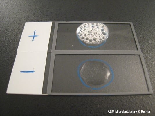
- Observe for the effervescence of gas formation that shows the organism has a catalase enzyme.
- Catalase producing organisms are: Staphylococcus aureus, Escherichia coli, Mycobacterium tuberculosis, Legionella pneumophila.

(Source:https://www.asmscience.org/content/education/imagegallery/image.3241 )
Oxidase test
- Oxidase test detects the bacteria that produce cytochrome C oxidase or cytochrome a3 when it undergoes an electron transport chain.
- The filter paper used is soaked with the reagent tetramethyl-p-phenylenediamine which develops dark blue-purple color when rubbed with an organism with oxidase enzyme giving positive results.
Procedure of Oxidase test
- A piece of filter paper soaked with 1% tetramethyl-p-phenylenediamine dihydrochloride is taken.
- The organism is rubbed on the paper with a sterile glass rod.
- Observe for the dark blue purple color formation within 30 second.
- Positive organisms are: Pseudomonas spp, Aeromonas spp, Vibrio spp, Neisseria gonorrhoeae.

Oxidative/Fermentative test
- O/F test determines whether an organism utilizes the substrate either in aerobic or anaerobic conditions.
- Two tubes are used in this method where one tube is open to the air and another is sealed with paraffin oil on the top.
- When an organism utilizes the substrate, it changes color due to the acid production that reacts with the pH indicator Bromo thymol blue.
Procedure of Oxidative/Fermentative test
- Take two tubes of O/F medium and label the organism.
- Stab the organism with a sterile inoculating wire.
- Put 1ml of paraffin oil in one tube only and incubate both tubes.
- Observe for the development of color from green to yellow which indicates the fermentation of substrate.
- Fermentative organisms: E. coli, S. cerevisiae.
- Oxidative organisms: P. aeruginosa.

IMViC test
Biochemical tests for bacteria identification
- IMVic test stands for Indole, Methyl Red, Voges Proskauer and Citrate tests which is done to separate coliforms.
- Indole test is done whether an organism produces the tryptophanase enzyme or not. If yes, then it hydrolyzes the tryptophan into indole.
- If the test organism gives a positive indole test, then it forms a cherry red color layer.
- The SIM (sulfur, Indole, Motility) medium is used in the indole test and Kovac’s reagent is also added as an indicator.
- It detects the motility and sulfide production.
Procedure of indole test
- Take the SIM media and inoculate the bacterial culture.
- After incubation, add 4-8 drops of Kovac’s reagent and mix the tube.
- Observe for the development of cherry red color, black precipitation for H2S production, and bacterial spread for motility.
- Indole positive organism is E. coli.
Methyl Red – Voges Proskauer (MR-VP) test
- This test is done to detect organisms ability to maintain stable acid or not as some organism undergoes mixed acid fermentation.
- Methyl red is added as a pH indicator to test the amount of acid which turns red at low pH which is a positive result and yellow at high pH as a negative result.
- Some species further produces more stable acids while some produces 2,3-butanediol as an end product which is detected by Voges Proskauer test.
- Barrit’s reagent A (α-Naphthol) and B (KOH) is added which gives red color.
Procedure of MRVP test
- Take two tube with MR-VP medium and inoculate with organism and incubate.
- Add 5-6 drops of MR reagent in one tube and Barrit’s reagent A and B in another and again incubate for few minutes.
- Observe for the red color formation.
- E. coli is a positive organism for MR whereas Enterobacter cloacae for VP.
Citrate test
- Some organism use citrate as a sole source of carbon for metabolism, those organisms can be detected by citrate test.
- Simmon’s citrate agar is used with Bromothymol blue as a pH indicator which gives prussian blue.
Procedure of Citrate test
- Take a slant of Simmon’s citrate agar and inoculate organism in a zigzag manner in a slat surface.
- Incubate and observe for color change which forms prussian blue indicates positive result.
- Positive organism: Klebsiella phenumomiae

Triple Sugar Iron Agar test (TSIA)
- TSIA test is done to detect the ability of organisms to ferment sugars and to reduce sulfites to sulfides which on production blackens the medium.
- Gas production can also be detected by the observation of crack and uplifting of the medium.
Procedure of Triple Sugar Iron Agar test (TSIA)
- Take a tube with TSIA and inoculate organism by stabbing at the butt and then streaking on the slant surface.
- Incubate and observe for the color change where yellow color indicates acid production whereas red color indicates alkali production in media, gas and H2S production.
- Possible organism: Citrobacter spp, E. coli, Shigella spp.

Urease test
This test detects the organism that produces urease enzyme and converts urea to ammonia and CO2 in the presence of water.
Phenol red is used as pH indicator which gives orange to deep pink color.
Procedure of Urease Test
- Take a tube of urea agar and streak the organism in the slant.
- Incubate and observe for color change.
- Positive organism: Proteus vulgaris

Nitrate reduction test
- This test detects the organism that produces nitrate reductase enzyme which reduces nitrate to nitrite.
- Nitrate reagent A and nitrate reagent B are used which develops red precipitation at the end of the test.
Procedure of Nitrate reduction test
- Take a nitrate broth and inoculate loopful of organism and incubate.
- Add 5 drops of nitrate reagent A and then 5 drops of B nitrate reagent and observe for develoment of red color.
- Nitrate reducing organisms: Neisseria mucosa, E. coli.

Learn more
Refrences:
- https://shodhganga.inflibnet.ac.in/bitstream/10603/131955/15/15_chapter%205.pdf
- https://www.slideshare.net/RaviKantAgrawal/biochemical-tests-for-identification-of-bacteria
- https://www.vetbact.org/index.php?biochemtest=1#id30
- http://www.uwyo.edu/molb2021/additional_info/summ_biochem/mrvp.html
- Manandhar.S, Sharma.S,(2013).Practical Approach to Microbiology, National Book Center,2nd edition (Page.65-81).