Blood vessels, the network of tubes through which blood is pumped, play a crucial role in the cardiovascular system. They work alongside the heart and blood to facilitate the circulation of blood throughout the body.
These channels form a closed loop, like a circuit, beginning and ending at the heart. With approximately 60,000 miles of intricate networks, blood vessels transport blood throughout the entire body. They serve as channels or conduits, distributing blood to body tissues. The circulatory system, consisting of the heart, blood cells, lymphatic system, and blood vessels, relies on these channels. The two closed systems of tubes, pulmonary vessels and systemic vessels, facilitate blood transportation between the heart, lungs, and body tissues.
Arteries, capillaries, and veins are the three main types of blood vessels, classified based on their structure and function. Understanding the significance of blood vessels is key to comprehending the circulatory system and its vital functions.
Anatomy of Arteries
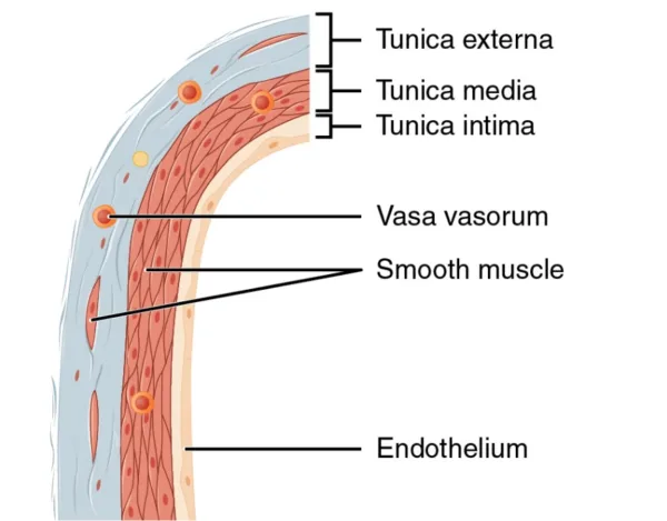
Arteries possess a distinct anatomy, consisting of three layers that contribute to their structure and function.
Tunica intima
The innermost layer, called the tunica intima or tunica interna, comprises a single layer of endothelial cells surrounded by a connective tissue basement membrane and elastic fibers. This layer forms a tube-like structure that allows oxygen-rich blood to flow through, preventing any leakage and ensuring targeted delivery of nutrients.
Tunica media
The middle layer, known as the tunica media, is predominantly composed of smooth muscle and is typically the thickest layer. It serves a dual purpose, providing structural support to the vessel and regulating blood flow and pressure by adjusting the diameter of the artery. The contraction of smooth muscles increases the pressure exerted on the arterial walls, while dilation reduces it during systolic pumping.
Tunica externa
The outermost layer, termed the tunica externa or tunica adventitia, connects the artery to surrounding tissues. It is primarily composed of connective tissue and contains varying amounts of elastic and collagenous fibers. This layer exhibits denser connective tissue near the tunica media and transitions to lose connective tissue towards the periphery of the vessel. The adventitia plays a crucial role in anchoring arteries and facilitating their connection to vascular nerves, which regulate the contraction of smooth muscles within the arteries.

The largest artery in the body is the aorta, which originates from the left ventricle of the heart. It branches into a network of smaller arteries that span throughout the body. Arterioles and capillaries are the smaller branches of arteries. Additionally, the pulmonary arteries are unique as they carry oxygen-poor blood from the heart to the lungs under low pressure.
Function of Arteries
Arteries transport pressurized blood away from the heart, typically carrying oxygenated blood except for the pulmonary artery, which conveys deoxygenated blood to the lungs.
- Arteries transport oxygen, nutrients, and hormones throughout the body.
- Oxygenated blood is carried by arteries to areas with low oxygen levels, where it is released through capillaries.
- Arteries adapt to signals from the central nervous system and external stimuli like pressure, temperature, and substances.
- Vascular nerves innervate arteries, enabling them to constrict or dilate, regulating blood pressure.
- Systemic arteries can be classified as muscular or elastic, with larger arteries being elastic and smaller ones being muscular, delivering blood to arterioles.
- Arteries are muscular-walled tubes connecting the heart and other body parts, capable of withstanding force and pressure.
- The aorta, the largest systemic artery, branches into smaller arteries throughout the body.
- Arteries carry oxygen-rich blood away from the heart, distributing it to different body parts.
- Two main types of arteries are pulmonary arteries, carrying blood from the heart to the lungs for oxygenation, and systemic arteries, supplying blood to the rest of the body.
- Pulmonary arteries facilitate the uptake of oxygen in the lungs, and oxygen-rich blood returns to the heart through pulmonary veins.
Anatomy of Veins
Veins, similar to arteries, consist of three layers that comprise their walls:
Tunica externa
This outer layer is the thickest and primarily composed of connective tissue. It houses tiny blood vessels called vasa vasorum, which supply blood to the vein walls.
Tunica media
The middle layer is thin and rich in collagen, a key component of connective tissue.
Tunica intima:
This innermost layer is a single layer of endothelial cells with some connective tissue. In certain veins, especially in the arms and legs, one-way valves are present within this layer to prevent blood from flowing backward.
Unlike arteries, veins have lower pressure and thinner, less elastic walls. This allows veins to hold a significant portion of circulating blood, exhibiting high capacitance. Veins accommodate a large volume of blood at lower pressures.
Additionally, the anatomy of veins includes the tunica adventitia, which is the outermost layer fused with surrounding tissue. The tunica media consists of collagen, elastic fibers, and smooth muscle fibers. The innermost layer, the tunica intima, is the thinnest and comprises an internal elastic membrane, connective tissue, and endothelium. The endothelium, in direct contact with blood flow, is the first to experience the effects of venous insufficiency.
The multi-layered structure of veins helps facilitate the return of blood towards the heart and maintain proper venous function in the circulatory system.
Functions of Veins
Veins collect deoxygenated blood from various body parts and transport it back to the heart. They are essential components of the circulatory system, working in coordination with other blood vessels and the heart to ensure continuous blood circulation.
- Transport deoxygenated blood back to the heart.
- Work in conjunction with arteries for blood circulation.
- Valves prevent backward blood flow, ensuring unidirectional flow.
- Accommodate larger blood volume due to low pressure.
- Act as blood reservoirs, storing significant amounts of blood.
- Aid in regulating body temperature by redistributing heat.
- Return metabolic waste products to the heart for elimination.
- Absorb and transport nutrients, hormones, and substances.
- Assist in maintaining blood pressure and adjusting blood volume.
- Support lymphatic system by draining excess fluid.
- Aid in returning blood from extremities against gravity.
- Enhance venous return through muscle contractions and body position changes.
Anatomy of Capillaries
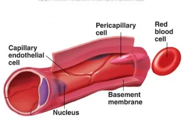
Capillaries, often referred to as hairlike vessels due to their small size, play a crucial role in the circulatory system. Here’s a breakdown of the anatomy of capillaries:
The structure of capillaries is uniquely designed to support their function. Comprised of a single layer of flattened endothelial cells without muscular or adventitial layers, capillaries are incredibly thin. This thinness enables close proximity to surrounding tissues, promoting efficient exchange. Nutrient and metabolite exchange primarily occurs through diffusion across the capillary walls. Blood flow through the capillaries is regulated by the arteriolar lumen, ensuring proper distribution and perfusion.
Capillaries have extremely thin walls, with a diameter of approximately 5 micrometers, consisting of only two layers of cells—an inner layer of endothelial cells and an outer layer of epithelial cells. Surrounding the capillary is the basement membrane, providing structural support and stability.
The human body houses an estimated 40 billion capillaries, which, if lined up in a single file, would stretch over 100,000 miles. These capillaries collectively form the capillary bed, an intricate network that supplies organs with the necessary nutrients and oxygen. The size and density of the capillary bed vary depending on the metabolic activity of the organ’s cells.
The capillary bed comprises true capillaries branching from arterioles and vascular shunts that create direct bypass pathways connecting arterioles and venules. Capillary width is passively regulated, as capillaries lack smooth muscle. Signaling molecules released by capillaries can affect the behavior of larger blood vessels in their vicinity.
During the inflammatory response, cytokines released can increase capillary permeability, allowing for enhanced immune cell access to affected areas. Additionally, the processes of vasculogenesis and angiogenesis play vital roles in embryological development, promoting the formation of new capillaries to ensure proper blood supply to developing tissues and organs.
Functions of Capillaries
Capillaries link the arterial system, encompassing blood vessels that transport blood away from the heart, with the venous system.
- Capillaries facilitate the exchange of nutrients, waste products, and interstitial fluid between the blood and tissue cells.
- They enable the diffusion of gases, such as oxygen and carbon dioxide, across their thin walls.
- Capillaries allow for the delivery of oxygen and nutrients to cells and the removal of metabolic waste products.
- They support the transport of hormones, antibodies, and other important substances throughout the body.
- Capillaries play a vital role in maintaining fluid balance by regulating the movement of fluids between the blood and surrounding tissues.
- They contribute to the regulation of blood pressure by controlling the resistance to blood flow.
- Capillaries provide a platform for immune responses, allowing white blood cells to migrate to infected or damaged tissues.
- They promote heat exchange by facilitating the transfer of heat from the blood to the surrounding tissues.
- Capillaries aid in the formation of new blood vessels through the processes of vasculogenesis and angiogenesis.
- They contribute to the regulation of local blood flow and distribution of blood to different tissues and organs.
Differences Between Arteries, Veins, and Capillaries
| Aspect | Arteries | Veins | Capillaries |
| Structure | Thick and elastic walls | Thinner and less elastic walls | Extremely thin walls |
| Layers | Three layers: tunica intima, tunica media, tunica externa | Three layers: tunica intima, tunica media, tunica externa | Single layer of endothelial cells |
| Blood Flow | Carry oxygenated blood except for pulmonary artery | Carry deoxygenated blood except for pulmonary veins | Facilitate exchange of nutrients and waste products |
| Pressure | High pressure | Low pressure | Low pressure |
| Valves | No valves | Valves present to prevent backward blood flow | No valves |
| Functions | Transport blood away from the heart | Transport blood towards the heart | Facilitate exchange of substances with tissue cells |
| Size | Vary in size from large to small arteries | Vary in size from large to small veins | Smallest blood vessels in diameter |
| Distribution | Branch into arterioles and capillaries | Merge into venules and then into veins | Connect arterioles to venules |
| Number | Fewer in number compared to capillaries and veins | More abundant compared to arteries | Most abundant and numerous in the circulatory system |
| Blood Volume | Carry a smaller volume of blood | Carry a larger volume of blood | Carry a small amount of blood due to their small size |
| Elasticity | Highly elastic | Less elastic | Not elastic |
| Direction of Blood Flow | Away from the heart | Towards the heart | Between arteries and veins |
| Oxygenation of Blood | Mostly oxygenated, except for pulmonary artery | Deoxygenated, except for pulmonary veins | Facilitate gas exchange, allowing both oxygenated and deoxygenated blood |
| Network Connection | Connected to arterioles and capillaries | Connected to venules and capillaries | Connect arteries and veins |
| Regulation of Blood Flow | Can constrict and dilate to regulate blood flow and pressure | Valves help prevent backflow of blood | No active control of blood flow |
FAQS
What are blood vessels, and what role do they play in the cardiovascular system?
Blood vessels are a network of tubes through which blood is pumped, and they facilitate the circulation of blood throughout the body. They work alongside the heart and blood to distribute blood to body tissues.
How are blood vessels classified?
Blood vessels are classified into three main types: arteries, capillaries, and veins. These classifications are based on their structure and function.
What is the anatomy of arteries?
Arteries have three layers: tunica intima, tunica media, and tunica externa. These layers contribute to their structure and function. The tunica intima is the innermost layer, followed by the tunica media, and the outermost layer is the tunica externa.
What are the functions of arteries?
Arteries transport oxygen, nutrients, and hormones throughout the body. They adapt to signals from the central nervous system and external stimuli. Arteries also regulate blood pressure through constriction and dilation.
How do veins differ from arteries?
Veins have thinner and less elastic walls compared to arteries. They transport deoxygenated blood back to the heart and have valves to prevent backward blood flow. Veins also serve as blood reservoirs and aid in maintaining blood pressure.
What is the anatomy of capillaries?
Capillaries are extremely thin-walled vessels consisting of a single layer of endothelial cells. They lack muscular or adventitial layers and facilitate the exchange of nutrients, waste products, and interstitial fluid between the blood and tissue cells.
What are the functions of capillaries?
Capillaries enable the diffusion of gases, such as oxygen and carbon dioxide, across their thin walls. They deliver oxygen and nutrients to cells, remove metabolic waste products, regulate fluid balance, and contribute to immune responses.
What are the key differences between arteries, veins, and capillaries?
Arteries have thick and elastic walls, carry oxygenated blood (except for the pulmonary artery), and transport blood away from the heart. Veins have thinner walls, carry deoxygenated blood (except for the pulmonary veins), and transport blood towards the heart. Capillaries have extremely thin walls, facilitate exchange between the blood and tissue cells, and have a single layer of endothelial cells.
How do arteries, veins, and capillaries differ in terms of pressure?
Arteries have high pressure, veins have low pressure, and capillaries have low pressure due to their thin walls.
What is the significance of blood vessels in the circulatory system?
Blood vessels are essential for the circulation of blood, delivering oxygen, nutrients, and hormones to body tissues while removing waste products. They regulate blood flow, maintain blood pressure, and support overall body function.
Learn more:
References
- American Heart Association. (2021). Anatomy of Blood Vessels.
- Cardiovascular System. (n.d.). Kenhub. Retrieved from https://www.kenhub.com/en/library/anatomy/the-cardiovascular-system
- Tortora, G. J., & Derrickson, B. (2017). Principles of Anatomy and Physiology (15th ed.). John Wiley & Sons.
- Waugh, A., & Grant, A. (2013). Anatomy and Physiology in Health and Illness (12th ed.). Elsevier Health Sciences.
- Britannica. (n.d.). Blood Vessel.
- Innerbody. (2023). Anatomy of Blood Vessels. Retrieved from