Lentiviral transduction is a widely used method for stable genetic modification of human induced pluripotent stem cells, or iPSCs. It is commonly used to introduce reporter genes, inducible transcription factors, shRNAs, miRNAs, CRISPR components, lineage-tracing systems, and selectable markers.
A common design is a lentiviral vector carrying GFP and puromycin resistance, either as separate expression cassettes or linked by an IRES or 2A peptide. GFP allows early visual or flow-cytometric monitoring of transduction efficiency, while puromycin enables enrichment or stable selection of transduced cells.
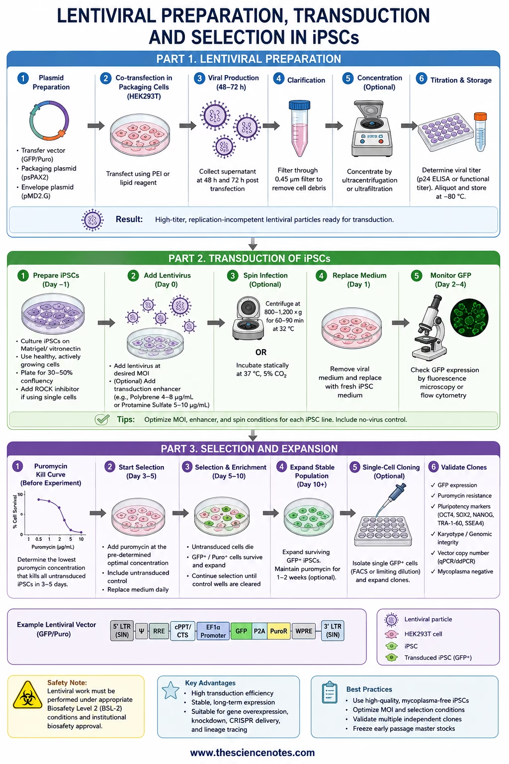
This protocol describes a research-use workflow for iPSC lentiviral transduction with GFP/puro selection, including culture preparation, transduction optimization, optional spin infection, puromycin kill curve, GFP-based assessment, antibiotic selection, clonal isolation, and quality control.
Scope and Biosafety
This protocol assumes that replication-incompetent, self-inactivating lentiviral particles have already been produced, concentrated if needed, titered, released for use, and approved under your institutional biosafety workflow.
Do not perform lentiviral work without:
- Institutional biosafety approval
- Lentiviral training
- Certified Class II biosafety cabinet
- Approved PPE
- Validated disinfectant and contact time
- Approved lentiviral waste handling
- Sealed centrifuge carriers or sealed rotors if spin infection is used
- Documented exposure-response procedure
Self-inactivating lentiviral systems are preferred for iPSC work, but SIN design does not eliminate the need for biosafety controls or downstream genomic validation.
Overview of the Workflow
| Day | Step |
|---|---|
| Before experiment | Confirm vector map, titer, GFP/puro design, and biosafety approval |
| Before experiment | Perform puromycin kill curve on parental iPSCs |
| Day −2 to −1 | Prepare healthy iPSCs and coated plates |
| Day −1 | Plate iPSCs for transduction |
| Day 0 | Add GFP/puro lentivirus ± enhancer ± spin infection |
| Day 1 | Replace virus-containing medium |
| Day 2–4 | Assess GFP expression and cell health |
| Day 3–5 | Begin puromycin selection after recovery |
| Day 5–14 | Continue selection and expand surviving GFP-positive population |
| Week 2–4 | Expand bulk population or isolate clones |
| Week 3+ | Validate GFP, puromycin resistance, pluripotency, genomic integrity, and vector copy number |
Materials
Cells and culture reagents
- Human iPSC line, mycoplasma-negative
- Validated iPSC maintenance medium, such as mTeSR, Essential 8, StemFlex, or lab-standard medium
- Validated matrix, such as Matrigel, vitronectin, laminin-521, or lab-standard substrate
- ROCK inhibitor, commonly used during single-cell plating or stress recovery
- Gentle dissociation reagent, such as EDTA-based reagent or Accutase, depending on the line
- DPBS or equivalent wash buffer
- Standard iPSC freezing medium
Lentiviral reagents
- Titered self-inactivating GFP/puro lentiviral stock
- Optional transduction enhancer:
- Protamine sulfate
- Polybrene
- Poloxamer-based enhancer
- LentiBOOST-type reagent
- Puromycin stock solution
- No-virus control
- Enhancer-only control
- Positive-control GFP lentivirus, if available
Assay reagents
- Flow cytometry buffer
- DAPI, PI, or other viability dye if flow cytometry is used
- Antibodies for pluripotency markers:
- OCT4
- SOX2
- NANOG
- TRA-1-60
- SSEA4
- qPCR or ddPCR reagents for vector copy number
- Mycoplasma test kit
- Karyotype, SNP array, or equivalent genomic integrity assay
Experimental Design
Do not run a single-condition transduction as the first attempt. iPSC lines differ in survival, transduction efficiency, promoter silencing, puromycin sensitivity, and clonal recovery.
Recommended pilot matrix:
| Condition | Virus | Enhancer | Spin infection | Purpose |
| Untransduced | No | No | No | Baseline morphology |
| Puromycin-control parental cells | No | No | No | Confirms kill curve |
| Enhancer-only | No | Yes | No | Enhancer toxicity |
| Low-virus static | Low | Optional | No | Low-copy candidate |
| Medium-virus static | Medium | Optional | No | Main condition |
| High-virus static | High | Optional | No | Maximum expression comparison |
| Low/medium-virus spin | Low or medium | Optional | Yes | Tests spin benefit |
| High-virus spin | High | Optional | Yes | High-efficiency but higher-stress comparison |
The best condition is not necessarily the one with the brightest GFP. The best condition is the one with acceptable GFP positivity, good viability, preserved iPSC morphology, manageable vector copy number, and good downstream differentiation performance.
Puromycin Kill Curve
Perform a kill curve before using puromycin selection. Do not copy puromycin doses from HEK293T cells, fibroblasts, or another iPSC line.
Purpose
To identify the lowest puromycin concentration that reliably kills untransduced parental iPSCs within the desired selection window.
Procedure
- Plate parental iPSCs in the same format, medium, matrix, and density planned for transduction.
- Allow cells to attach and recover overnight.
- Apply a puromycin concentration series.
- Include a no-puromycin control.
- Feed cells on the same schedule planned for the actual experiment.
- Monitor daily for:
- Cell death
- Detachment
- Surviving colonies
- Differentiation
- Morphologic stress
- Select the lowest puromycin concentration that eliminates all untransduced iPSCs within the intended selection period.
- Repeat the kill curve if the iPSC line, medium, matrix, passage method, or culture format changes.
Practical interpretation
- If all cells die too quickly, the dose may be unnecessarily harsh.
- If parental cells survive too long, the dose may be too weak.
- For iPSCs, excessive antibiotic stress can select for abnormal or unusually resistant subpopulations, so use the minimum effective dose.
Day −1: Plate iPSCs for Transduction
Goal
Plate iPSCs so that they are healthy, attached, and sub-confluent on the day of transduction.
Procedure
- Coat culture plates using the validated matrix for the iPSC line.
- Prepare iPSCs from a healthy, low-differentiation culture.
- Dissociate cells using the lab’s standard method:
- Small clumps for fragile lines
- Single cells for more uniform lentiviral exposure
- Plate cells in complete iPSC medium.
- Add ROCK inhibitor if using single-cell plating or if the line is stress-sensitive.
- Incubate overnight under standard iPSC culture conditions.
Target state on Day 0
Cells should be:
- Attached
- Evenly distributed
- Actively proliferating
- Not over-confluent
- Low in spontaneous differentiation
- Healthy by morphology
If cells are sparse, stressed, or differentiating on Day 0, delay transduction rather than proceeding with a compromised culture.
Day 0: GFP/Puro Lentiviral Transduction
Static transduction procedure
- Inspect cells by phase-contrast microscopy.
- Exclude wells with poor attachment, excessive differentiation, or uneven density.
- Thaw lentiviral aliquot according to your institutional SOP.
- Prepare virus-containing iPSC medium for each test condition.
- Add transduction enhancer only if it is part of the optimization matrix.
- Remove spent medium from iPSCs.
- Add virus-containing medium gently.
- Return cells to the incubator.
- Record:
- iPSC line
- Passage number
- Matrix
- Medium
- Cell density
- Vector name
- Vector lot
- Functional titer
- Virus amount or target MOI
- Enhancer and concentration
- Exposure duration
- Operator
- Date
Optional spin infection
Spin infection should not be the default for iPSCs. Use it only as a tested optimization condition and only if allowed by your institutional lentiviral SOP.
Spin infection may improve virus-cell contact, but it can also increase stress, detachment, and differentiation. Include a no-spin condition for comparison.
Evaluate spin infection by comparing:
- GFP-positive fraction
- Mean GFP intensity
- Cell survival
- Detachment
- Differentiation
- Recovery after medium change
- Vector copy number
- Pluripotency marker retention
If spin infection improves GFP but worsens survival or pluripotency, do not use it for line generation.
Day 1: Medium Replacement
- Remove virus-containing medium according to approved lentiviral waste procedures.
- Replace with fresh complete iPSC medium.
- Continue ROCK inhibitor briefly if cells were single-cell plated or visibly stressed.
- Inspect morphology and survival.
- Record:
- Detachment
- Cell death
- Colony morphology
- Differentiation
- Confluency
Avoid harsh washing if the iPSC line detaches easily. If washing is required by your SOP, use the gentlest validated approach.
Day 2–4: GFP Assessment
GFP expression is usually assessed after the vector has had time to express. The exact timing depends on promoter, vector design, and iPSC recovery.
Qualitative GFP check
Use fluorescence microscopy to estimate:
- Whether GFP is detectable
- Whether expression is uniform or mosaic
- Whether GFP-positive cells retain iPSC morphology
- Whether bright GFP correlates with cell stress
- Whether GFP is present in differentiated cells, undifferentiated colonies, or both
Quantitative GFP check
Flow cytometry is recommended if the result will determine selection strategy.
Measure:
- Percentage GFP-positive cells
- Mean fluorescence intensity
- Viability
- Scatter profile
- Optional pluripotency marker co-expression
Suggested interpretation:
| Result | Meaning | Action |
| Low GFP, healthy cells | Under-transduced | Increase virus, test enhancer, or test spin condition |
| High GFP, high death | Too harsh | Reduce virus, remove enhancer, avoid spin |
| High GFP, healthy morphology | Candidate condition | Proceed to selection |
| GFP mainly in differentiated cells | Poor culture quality or marker bias | Improve iPSC culture before repeating |
| Mosaic GFP | Expected after bulk transduction | Use selection or clonal isolation |
Day 3–5: Start Puromycin Selection
Do not start puromycin immediately after transduction unless the system has already been validated. Starting too early can kill transduced cells before puromycin resistance is sufficiently expressed.
When to start
Begin selection when:
- Cells have recovered from transduction
- GFP is detectable
- Colonies are attached and viable
- The planned kill-curve dose is available
- No-virus selection control is included
Puromycin selection procedure
- Replace medium with fresh iPSC medium containing the kill-curve-defined puromycin concentration.
- Apply the same puromycin concentration to the untransduced control.
- Feed daily or according to puromycin stability and iPSC health.
- Monitor morphology and death daily.
- Continue selection until the untransduced control is fully eliminated.
- Maintain surviving transduced cells until they recover and expand.
- Reduce, pulse, or discontinue puromycin only if compatible with the vector design and experimental goal.
Selection endpoints
A successful selection should produce:
- Clear death of untransduced control cells
- Survival of transduced cells
- Enrichment of GFP-positive cells
- Retention of iPSC morphology
- Minimal spontaneous differentiation
If puromycin selection causes widespread death even in GFP-positive wells, the dose may be too high, selection may have started too early, or transgene expression may be insufficient.
GFP-Based Enrichment Versus Puromycin Selection
GFP and puromycin can be used separately or together.
GFP monitoring only
Use when:
- You need early transduction readout
- You plan to isolate clones manually
- Antibiotic selection is too stressful
- The vector is not intended for long-term antibiotic pressure
Puromycin selection only
Use when:
- GFP is absent or not reliable
- The marker is non-fluorescent
- Bulk enrichment is sufficient
GFP plus puromycin
Use when:
- You want early visual confirmation
- You need antibiotic enrichment
- You want to sort or pick GFP-positive colonies
- You want to confirm that puromycin-resistant cells are also GFP-positive
FACS sorting for GFP
FACS can rapidly enrich GFP-positive iPSCs, but it adds single-cell stress. If sorting is used:
- Use viability dye
- Use gentle dissociation
- Sort into supportive medium
- Plate onto optimized matrix
- Use ROCK inhibitor
- Avoid overly stringent gates if expression is still maturing
- Allow recovery before further selection or cloning
Bulk Expansion After Puromycin Selection
Bulk GFP/puro-selected populations are useful for pilot assays and some screening workflows.
Procedure
- Continue feeding selected cells until surviving colonies expand.
- Remove obvious differentiated regions manually if necessary.
- Passage gently before cultures become over-confluent.
- Maintain puromycin only if needed to preserve the transduced population.
- Confirm GFP enrichment by microscopy or flow cytometry.
- Freeze early-passage bulk stocks.
Limitations of bulk populations
Bulk populations may contain:
- Multiple integration sites
- Variable copy numbers
- Different GFP intensities
- Differentiation-prone subpopulations
- Partially silenced cells
- Mixed biological behavior
For disease modeling, differentiation phenotyping, rescue studies, or mechanistic experiments, clonal isolation is usually preferable.
Clonal Isolation of GFP/Puro iPSCs
Clonal iPSC lines are recommended when reproducibility, genotype, expression level, or differentiation phenotype matters.
Timing
Begin clonal isolation after:
- Cells survive puromycin selection
- GFP expression is detectable
- iPSC morphology is recovered
- Cultures are not over-stressed
Methods
Common options include:
- Limiting dilution
- Single-cell FACS sorting of GFP-positive cells
- Manual picking of GFP-positive colonies
- Semi-clonal low-density plating followed by colony picking
Clonal isolation procedure
- Prepare matrix-coated plates.
- Prepare supportive iPSC medium with ROCK inhibitor.
- Dissociate selected GFP-positive cells gently.
- Plate at clonal density or sort single GFP-positive cells.
- Avoid disturbing plates during early colony formation.
- Feed gently.
- Identify emerging colonies with good iPSC morphology.
- Confirm GFP expression by microscopy.
- Pick individual colonies into separate wells.
- Expand gradually.
- Freeze early backup vials.
- Validate before downstream use.
Clone selection criteria
Prioritize clones that show:
- Normal iPSC morphology
- Stable GFP expression
- Puromycin resistance
- Robust expansion
- Minimal differentiation
- Moderate expression, if overexpression toxicity is possible
- Acceptable vector copy number
- Normal genomic integrity
Avoid clones that show abnormal growth, excessive differentiation, unusually high GFP with poor morphology, or poor recovery after passaging.
Clone-Level Quality Control
1. GFP and puromycin validation
Confirm:
- GFP-positive percentage
- Mean GFP intensity
- GFP stability across passages
- Survival under puromycin
- Loss of parental cells in selection control
- No major GFP silencing after expansion
2. Vector confirmation
Recommended assays:
- PCR for vector-specific sequence
- qPCR or ddPCR for vector copy number
- RT-qPCR for transcript expression, if relevant
- Western blot or immunostaining for protein expression, if relevant
For many iPSC applications, moderate-copy clones are preferable to very high-copy clones.
3. Pluripotency testing
Validate:
- OCT4
- SOX2
- NANOG
- TRA-1-60
- SSEA4
- Normal compact colony morphology
Use immunostaining, flow cytometry, RT-qPCR, or a combination depending on the lab’s standard QC workflow.
4. Genomic integrity
Recommended:
- G-banded karyotype, SNP array, or equivalent assay
- Vector copy number
- Integration-site analysis if the application requires it
- Off-target analysis if CRISPR components were delivered
5. Functional validation
Before using clones in major experiments, confirm:
- Differentiation capacity
- Lineage-specific marker expression
- Expected phenotype
- Stability of GFP through differentiation, if required
- No unexpected growth or survival advantage
- Comparable behavior to parental and mock-treated controls
Troubleshooting GFP/Puro iPSC Transduction
Problem: GFP is low after transduction
Possible causes:
- Low functional titer
- Poor promoter activity in iPSCs
- Dense colonies
- Poor vector access
- Short exposure
- No enhancer
- Lentivirus lost activity after freeze-thaw
Solutions:
- Use fresh or higher-titer virus
- Test a higher virus condition
- Optimize plating density
- Compare single-cell versus clump plating
- Test protamine sulfate or another enhancer
- Test spin infection side by side
- Confirm promoter is active in iPSCs
Problem: GFP is high but cells die
Possible causes:
- Excess viral load
- Enhancer toxicity
- Spin infection stress
- Transgene toxicity
- Single-cell stress
- Poor starting culture
Solutions:
- Reduce virus dose
- Remove enhancer
- Avoid spin infection
- Use ROCK inhibitor support
- Delay selection
- Test inducible or weaker promoter design
- Improve iPSC culture before repeating
Problem: Puromycin kills all cells
Possible causes:
- Selection started too early
- Puromycin dose too high
- Resistance cassette not expressed well
- Vector silencing
- Poor transduction efficiency
- Cells were already stressed
Solutions:
- Delay selection until GFP is detectable and cells recover
- Repeat kill curve
- Lower puromycin concentration
- Extend recovery period
- Use GFP sorting instead of antibiotic selection
- Screen vector design
Problem: Puromycin selection leaves GFP-negative cells
Possible causes:
- Separate cassette behavior
- Low GFP expression threshold
- Promoter silencing of GFP but not puro
- Autofluorescence misinterpretation
- Mixed population
Solutions:
- Confirm by flow cytometry
- Re-sort GFP-positive cells
- Isolate clones
- Validate vector sequence and expression cassette
- Confirm puromycin resistance is vector-derived
Problem: GFP fades over passages
Possible causes:
- Promoter silencing
- Loss of selection
- Clone-specific integration effect
- Differentiation-associated silencing
Solutions:
- Maintain puromycin if appropriate
- Screen additional clones
- Use a more stable promoter
- Test expression after differentiation
- Avoid relying on CMV promoter unless validated in your system
Problem: Clones differentiate after selection
Possible causes:
- Overly harsh puromycin selection
- Spin infection stress
- Single-cell stress
- Poor matrix or medium
- Picking poor-quality colonies
Solutions:
- Reduce selection pressure
- Improve recovery conditions
- Use ROCK inhibitor during cloning
- Pick only high-quality colonies
- Avoid over-confluence
- Repeat from healthier starting iPSCs
Best Practices for GFP/Puro iPSC Lentiviral Transduction
- Always perform a puromycin kill curve.
- Use GFP to assess timing before starting selection.
- Do not start puromycin immediately unless validated.
- Include untransduced puromycin-control wells.
- Optimize virus dose rather than maximizing it.
- Treat spin infection as optional, not routine.
- Confirm that GFP-positive cells retain iPSC morphology.
- Expand multiple independent clones.
- Measure vector copy number when interpretation matters.
- Validate pluripotency and genomic integrity before differentiation studies.
- Freeze early-passage stocks.
Conclusion
GFP/puro lentiviral transduction is a practical and powerful strategy for generating stable genetically modified iPSC lines. GFP provides a real-time readout of transduction efficiency, while puromycin enables enrichment of transduced cells. However, successful iPSC lentiviral transduction requires more than GFP positivity and antibiotic survival.
The final goal is a healthy, genomically stable, pluripotent iPSC population or clone with reproducible expression and reliable differentiation performance. For that reason, every GFP/puro workflow should include a virus-dose optimization, puromycin kill curve, careful timing of selection, optional but controlled spin infection testing, clonal validation when needed, and rigorous QC before downstream experiments.