- The word ‘Microscope’ (plural – Microscopes) is derived from the Latin word ‘Microscopium’ where ‘mikros’ means small and ‘skopein’ meaning ‘to look at’.
What is a microscope?
- A microscope is an instrument that has a powerful magnifying resolution lens which enlarges the image that cannot be visualized by our naked eye and reveals the detail for further study and provides us an information.
- Discovery of microscope is still unclear but through the information of some historians, Hans Lippershey, who invented telescope in about 1590 seem to have made the concept of earliest microscopes.
- But Galileo Galilei improved the microscope device in early 16th century. Later, Anton Van Leuwenhoek perfected the lens resolution that increased the image size.
- Many discoveries and improvement of microscope had made by several scientists like August Kohler, Ernst Leitz and Ernst Abbe who were credited on inventing microscope photographs, different magnification in single microscope and ultraviolet microscope.
- Photographs can also be taken while using the microscope which is called as micrographs.
- The microscopic resolution is expressed in micrometers (µm).
- Biological scientists, microbiologists, biochemists and biophysicist frequently use microscope to study the behavior of microorganisms and for research purpose.
Microscopes are distinguished on the basis of magnification and resolution power:
What is Magnification?
Magnification is the measure of an object to appear large in a microscope. The common light microscope that we use in schools and colleges magnify up to 400 times the original size.
What is Resolution of a Microscope?
Resolution is a clear distinguishable power as two objects that might look single in a low resolution appears to be separated in high resolution. Smaller the value, higher will be the resolving power.
Light microscope
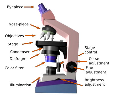
- Light microscope is also referred as an optical microscope.
- Light microscope uses a visible light and magnifies the specimen in finer detail.
- The basic principle of light microscopy is that when an image is magnified through an eyepiece, it passes through one or two lenses for magnification and finally an image is viewed.
- This microscope is usually used by the students and researcher in laboratories as it has benefits of observing living cells carrying out their behavior like dividing and migrating.
- Based on the number of lenses, light microscope is of two types:
- Simple light microscope (contain single lens)
- Compound light microscope (contain two lens)
Light microscope is further divided into four modern types:
Types of Microscopes
- Bright field light microscope
- Phase contrast light microscope
- Dark field light microscope
- Fluorescence light microscope
Bright field light microscope:
- Bright field microscopy image appears to be dark on a bright background. Therefore, its name is given according to the light and background it uses.
- A visible light when passes through the specimen forms an image directly without any modification. It’s simple working mechanism made this microscopy a popular technique.
- Bright field microscopy is also known as compound light microscopy and is widely used in laboratory to view animal and plant cells and bacteria and some parasites as well.
Phase contrast microscope:
- Phase contrast microscope converts the phase shifts when the light passes through the cell which is translucent i.e., unstained.
- By using a high-resolution objective lens, these phase shift gets converted into an amplitude, but it requires a specialized and expensive condenser and objective lens.
- PCM views the unstained cells because staining and fixing kills most of the cells and the morphology will not be clearly understood and also disturbs the cell natural state.
- PCM applies in the study of motility and structure of microorganisms. It also determines the morphology of animal and plant cells and detects the bacterial endospores.
Dark-field light microscope:
- The image when focused on the dark field light microscope appears to be illuminated while the background appears to be dark.
- Basic principle is that when a beam of light gets transmitted to an object, an unreflected rays do not pass through the objectives. Therefore, the background appears to be dark.
- It visualizes the unstained living cells and the internal organs of eukaryotic cells. It is also used in identification of bacterial cells with its distinctive shape and sizes.
Fluorescent microscope:
- Fluorescent microscope is another type of light microscope which differs from other light microscopes.
- Other microscope provides an image only after the transmission of light, but fluorescence microscopy absorbs one wavelength of light and emits another.
- One wavelength of light excites the fluorescent molecules, and the other different wavelength is collected and forms a picture known as fluorochrome labelled image.
- The four main types of light source that the fluorescence microscope uses are Xenon arc lamp, lasers, supercontinuum source and high-power LEDs.
- In most of the cases, the cells or tissue we desire to study is not naturally fluorescent so we must label it with a fluorescent dye before observation.
- Dyes like Rhodamine B and Fluorescein-5-isothiocenate (FITC) are used for the visualization and detection of proteins, nucleic acids, antigens/pathogens and other cellular components.
Electron microscope
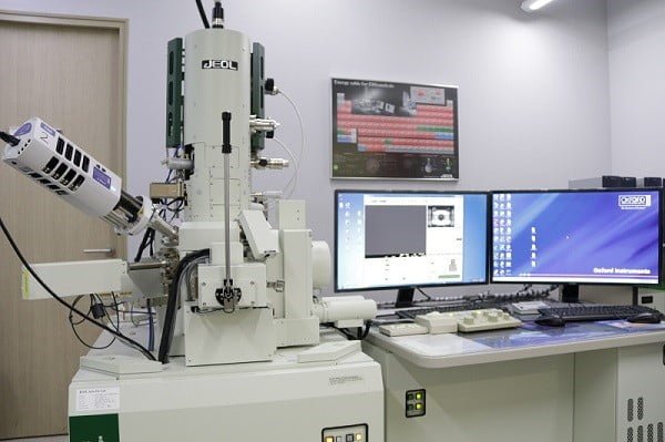
- Electron microscopes produces much higher resolution image than the light microscope that we studied earlier.
- They magnify the image in nanometers because they use a beam of electrons to visualize a specimen rather than a beam of light.
- Electron microscope has made a mark of progress in the technology, all thanks to Max Knoll and Ernst Ruska who finally overcome the barrier and limitations of visible light in 1931 at the Berlin Tekniche Hochschule.
- Electron microscope is used to examine the subcellular structure and compartment of cells, tissue and organelles. It is also used in biomedical research to investigate the disease and also in the development and manufacturing of electronics.
On the basis of different operating style, electron microscope is of two Types of Microscopes
- Scanning electron microscope (SEM)
- Transmission electron microscope (TEM)
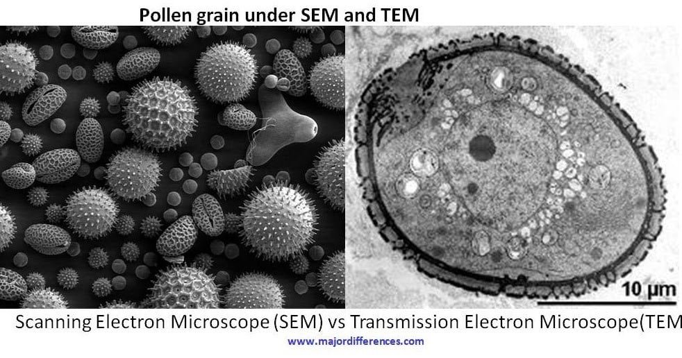
Scanning electron microscope:
- In SEM, a beam of electrons is scanned back and forth across the surface of the cell which creates a 3D image.
- It depends upon the emission of secondary electrons.
- It has a high tension of ~1-30 kV and maximum magnification up to 1-2million times.
- It is easy to use and require no sample preparation.
Transmission electron microscope:
- TEM uses thin specimen <150nm where parallel or sliced beam of electron passes over its surface.
- It has high tension of ~60-300 kV and magnifies more than 50 million times.
- It can only be used by trained personnel and requires laborious sample preparation.
Electron Microscopy: Types, Instrumentation, Principle, and Applications (thesciencenotes.com)
References:
- https://www.britannica.com/technology/microscope
- Houck, M. M., & Siegel, J. A. (2010). Microscopy. Fundamentals of Forensic Science, 77–97.
- https://www.khanacademy.org/science/high-school-biology/hs-cells/hs-introduction-to-cells/a/microscopy
- https://microbenotes.com/electron-microscope-principle-types-components-applications-advantages-limitations/