In the era of rising antibiotic resistance, the “one size fits all” approach to antimicrobial therapy is no longer sufficient. To save lives in critical clinical settings—such as cases of bacterial endocarditis or sepsis—physicians need to know exactly how much of a drug is required to stop a specific pathogen. This is where the Minimum Inhibitory Concentration (MIC) comes into play.
This article provides an in-depth educational look at what MIC is, why it is essential for modern medicine, and the gold-standard laboratory methods used to determine it, including Broth Dilution, Agar Dilution, and the E-test.
Minimum Inhibitory Concentration (MIC) is defined as the lowest concentration of an antimicrobial agent that inhibits the visible growth of a microorganism after overnight incubation.
Unlike qualitative tests (like the Kirby-Bauer disk diffusion method) that simply tell you if a bacterium is “Sensitive” or “Resistant,” the MIC provides a quantitative value. It is usually measured in mcg/mL (micrograms per milliliter) or mg/L (milligrams per liter). This number represents the exact potency of the antibiotic against a specific strain of bacteria.
MIC vs. MBC: What’s the Difference?
While MIC measures the concentration needed to inhibit growth (bacteriostatic), the Minimum Bactericidal Concentration (MBC) measures the lowest concentration required to actually kill the bacteria. Determining the MBC is often a secondary step following an MIC test.
Why is MIC Testing Crucial in Healthcare?
Estimating the MIC is not just a laboratory exercise; it has profound implications for patient outcomes.
1. Regulating Therapeutic Doses
In life-threatening situations like bacterial endocarditis, where the infection sits on heart valves, dosing must be incredibly precise. The MIC allows clinicians to calculate a dose that is high enough to be effective but low enough to avoid systemic toxicity to the patient’s organs (like the kidneys or liver).
2. Testing Slow-Growing Bacteria
Some bacteria, such as Mycobacterium tuberculosis, grow very slowly. Traditional sensitivity tests are difficult to interpret with these organisms. MIC testing provides a reliable quantitative pattern of antimicrobial sensitivity for these complex cases.
3. Monitoring Resistance Trends
By tracking MIC values over time, public health officials can see if a specific bacterial population is becoming “less sensitive” to an antibiotic, even before full-blown resistance develops.
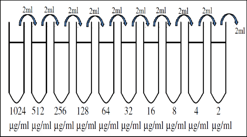
Schematic of the double dilution method: This diagram illustrates the serial “doubling” dilution process, showing the transition from visible bacterial turbidity to clear tubes that represent the Minimum Inhibitory Concentration (MIC).
Method 1: The Broth Dilution Method
Microbiologists favor the Broth Dilution method as a classic, quantitative in vitro technique. This approach tests a specific organism against decreasing concentrations of an antibiotic within a liquid medium to find the exact point of growth inhibition.
The Procedure
To perform this test, follow these standard laboratory steps:
Serial Dilution: Serially dilute the antimicrobial agent in Mueller–Hinton broth. Most labs utilize a “doubling dilution” technique (for example: 64 mcg/mL, 32 mcg/mL, 16 mcg/mL, 8 mcg/mL, etc.).
Inoculation: Add a standard suspension of the test organism’s broth culture to each antibiotic dilution and a designated control tube.
Controls: Include an organism with known susceptibility as a control. This ensures that both the drug and the broth media perform correctly.
Incubation: Mix the tubes gently and incubate them at 37°C for 16–18 hours.
Interpreting Results
Record the MIC by identifying the lowest concentration of the drug that shows no visible growth. In the laboratory, technicians identify growth by looking for turbidity (cloudiness) in the tube. If the tube remains clear, the antibiotic has successfully inhibited the bacteria.
Determining the MBC (Minimum Bactericidal Concentration)
The broth method offers a significant advantage: you can determine the MBC using the same samples from the MIC test.
Perform subcultures by transferring samples from each tube showing no growth onto nutrient agar plates that contain no antibiotics.
Examine the plates for any bacterial growth after incubating them overnight at 37°C.
Identify the MBC by finding the tube with the lowest drug concentration that fails to produce any growth on the subculture plate.
Pro Tip:Broth microdilution is performed using microtiter plates. Because it is efficient and precise, it is considered the “gold standard” in modern microbiology.
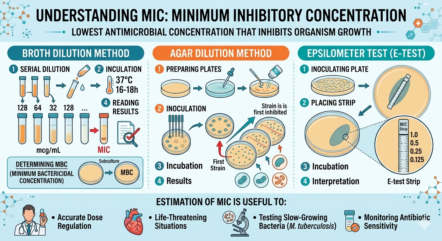
A visual guide to MIC determination: This infographic illustrates the laboratory procedures for Broth Dilution, Agar Dilution, and the Epsilometer test (E-test). It highlights how these quantitative methods help clinicians regulate antibiotic doses for life-threatening conditions and monitor the sensitivity of slow-growing bacteria like M. tuberculosis.
Method 2: The Agar Dilution Method
The Agar Dilution method is another quantitative approach, but it utilizes a solid medium rather than a liquid one.
The Procedure
To ensure accuracy and reliability during agar dilution, follow these structured steps:
Preparation: Create serial dilutions of the antibiotic using distilled water as the diluent.
Incorporation: Add these antibiotic dilutions to Mueller–Hinton agar that you have melted and cooled to a maximum of 60°C. Pour the resulting mixture into Petri dishes to solidify.
Control Plate: Inoculate one control plate without any antibiotics. This step confirms the viability of the organism and ensures it grows normally under test conditions.
Inoculation: Inoculate the test organism onto the prepared plates and incubate them overnight at 37°C.
Interpreting Results
After the incubation period, examine the plates for the presence or absence of bacterial growth:
Identify the MIC: Note the lowest concentration of the antibiotic that completely inhibits bacterial growth; this value represents the MIC.
Classification: Compare your MIC results with established CLSI (Clinical and Laboratory Standards Institute) guidelines. Report the organisms as Sensitive, Intermediate, or Resistant based on these standards.
Main Advantage: This method allows you to test a large number of different organisms simultaneously on a single plate. This high-throughput capability makes it exceptionally efficient for large-scale research studies and clinical trials.
Method 3: The Epsilometer Test (E-test)
The E-test is an automated-style system for measuring the MIC of a bacterial isolate. It combines the principles of disk diffusion with the quantitative accuracy of dilution methods.
How it Works
The E-test uses an absorbent plastic strip:
One Side: Contains a continuous gradient of the antibiotic immobilized on the surface.
Other Side: Contains an MIC interpretative scale corresponding to 15 twofold MIC dilutions.
The Procedure
The strip is placed on an agar plate previously inoculated with the test organism.
The MIC scale must face toward the opening side of the plate.
During incubation at 37°C overnight, the antibiotic diffuses into the agar.
An elliptical zone of inhibition (shaped like a teardrop) forms around the strip.
Reading the E-test
The MIC value is read directly from the scale at the intersection of the inhibition zone with the strip.
Important: The end point must always be read at the point of complete inhibition of all growth, including any faint hazes or isolated micro-colonies. The E-test is highly valued because it is very easy for laboratory technicians to interpret.
Comparison of MIC Determination Methods
Feature
Broth Dilution
Agar Dilution
E-Test
Medium
Mueller–Hinton Broth
Mueller–Hinton Agar
Mueller–Hinton Agar
Efficiency
Best for few isolates
Best for many isolates
One strip per drug
Bactericidal Data
Can determine MBC
No MBC data
No MBC data
Setup Complexity
High (Serial dilutions)
Moderate
Low (Ready-to-use)
Result Type
Quantitative
Quantitative
Quantitative
Key Factors for Accurate MIC Testing
To ensure results are Google-friendly and scientifically accurate, laboratories must adhere to strict standards:
Temperature: Must be maintained at 37°C; deviations can lead to false resistance or sensitivity.
Media Quality: Mueller–Hinton media is the standard because it allows for even diffusion and lacks common antibiotic inhibitors.
Inoculum Standardization: The concentration of bacteria added to the test must be consistent (usually matched to a 0.5 McFarland standard).
Conclusion
Understanding and determining the Minimum Inhibitory Concentration (MIC) is a cornerstone of modern infectious disease management. Whether using the manual Broth Dilution method to find the MBC, the high-volume Agar Dilution method, or the user-friendly E-test, these tools allow scientists and doctors to fight bacterial infections with surgical precision.
As antibiotic resistance continues to challenge global health, the quantitative data provided by MIC testing will remain our most effective weapon in ensuring successful patient recovery.
Alisha G C is an MBBS student at Nepalgunj Medical College, Banke, Nepal. She writes biology notes at www.thesciencenotes.com.
https://www.nature.com/articles/d41586-025-00589-z
View all posts by Alisha G C